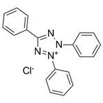
2,3,5-Triphenyltetrazolium Chloride: A Versatile Dye for Tissue Vitality Detection and Ablated Area Evaluation
2,3,5-Triphenyltetrazolium chloride is a colorless water-soluble dye. In the mitochondria of living cells, it is reduced to a deep red, water-insoluble compound (formazan). It helps to distinguish between viable and infarcted brain tissue after stroke. 2,3,5-Triphenyltetrazolium chloride is a colorless water-soluble dye. In the mitochondria of living cells, it is reduced to a deep red, water-insoluble compound (formazan). 2,3,5-Triphenyltetrazolium chloride helps to distinguish between viable and infarcted brain tissue after stroke.

Evaluation of electroporated area using 2,3,5-triphenyltetrazolium chloride
Magnetic resonance imaging (MRI) was used for instant imaging of potato tissue changes after pulsing to overcome these disadvantages. The ablation area detected using MRI was similar to the conventional melanin pigmentation ablation outcome in various electric field strengths. However, using MRI for most researchers is unsuitable due to its high cost and uneasiness to handle. The water-soluble tetrazolium salt has been used to evaluate cell viability in in vitro and ex vivo experiments. The 2,3,5-triphenyltetrazolium chloride (TTC) is a representative agent for this purpose and is reduced to insoluble red-colored triphenylformazan crystals by the dehydrogenase produced by mitochondria in cells. The IRE-ablated area was evaluated with TTC in ex vivo swine pancreatic tissue. The TTC-unstained white area was an IRE-ablated area in the swine pancreas tissue. This type of TTC staining has a rapid chemical reaction and reduces evaluation time. However, research is needed to evaluate the IRE-ablated area potato models using TTC staining. Here, an IRE-ablated area was assessed based on 2,3,5-Triphenyltetrazolium chloride staining in a potato model after pulsing. The TTC-unstained white area was compared with the blackened area by the conventional melanin accumulation method.[1]
The recommendation is that experimental time should be short when using potato tissues after pulsing because living tissues, like potatoes, are susceptible to the external environment, such as ambient temperature and humidity. Moreover, the consistency of the IRE-ablated area using 2,3,5-Triphenyltetrazolium chloride staining was verified at different staining times in the present study experiments. The limitation of this study was that the TTC-stained presumed-RE area was not directly identified. Additional microscopy studies are needed to investigate whether the potato cells in the presumed-RE region show reversible nanopores after pulsing. Furthermore, the mechanism of this phenomenon should be investigated in future studies. In summary, the IRE-ablated area was distinguished with 2,3,5-Triphenyltetrazolium chloride staining in a potato model within 3 h and showed results similar to conventional melanin accumulation area after pulsing. The TTC-unstained white areas were consistent over time and at different staining times. The presumed-RE area was formed with TTC staining in a potato model. The areas were highly correlated with electrical variables such as current and conductivity changes. Hence, 2,3,5-Triphenyltetrazolium chloride staining might be the best choice to evaluate IRE-ablated and RE areas in a potato model.
Mannitol-facilitated perfusion staining with 2, 3, 5-triphenyltetrazolium chloride
Immersion staining of fresh brain slices with 2,3,5-triphenyltetrazolium chloride (TTC) is a simple and popular method for detecting infarction in experimental stroke models. TTC, a colorless water-soluble dye, is reduced to a deep red, water-insoluble compound (formazan) predominantly in the mitochondria of living cells, hence distinguishing between viable and infarcted brain tissue after stroke. However, immersion TTC-staining has its limitations because it is often difficult to section unfixed, edematous brains after severe ischemic injury, especially in newborn rodents. While perfusion staining of rodent brains by intracardiac injection of TTC solution was reported, we have found this method to be very inefficient (see Results), and the previous 2,3,5-Triphenyltetrazolium chloride perfusion-labeling method was rarely adopted in the literature. The lack of a reliable in-vivo TTC-labeling method has increased the workload of quantifying tissue damage in experimental stroke, and uncertainty in distinguishing the core-versus-peri infarct areas for biochemical analysis. Interestingly, although intracardiac injection of TTC hardly labels the brain, it stains the heart efficiently and has been used widely to detect experimental myocardial infarction. This disparity suggests to us that the entry of TTC dye to central nervous system may be hindered by the blood-brain-barrier (BBB). To test this hypothesis, we performed TTC perfusion-labeling after osmotic BBB disruption with mannitol, and found a greatly enhanced brain-staining capacity. Here we report optimization of the 2,3,5-Triphenyltetrazolium chloride perfusion-labeling method that demarcates infarction in both adult and newborn mouse brains.[2]
In conclusion, our results demonstrated a mannitol-facilitated 2,3,5-Triphenyltetrazolium chloride perfusion-labeling method that is a useful alternative to the ex-vivo immersion-labeling method. The in-vivo TTC-labeling method is simple, inexpensive, and particular useful for measuring injury in severely damaged or small newborn brains. This new vitality-detection method not only simplifies the task of quantifying infarction, but also supports biochemical analysis of the core- and peri-infarct areas in experimental stroke.
References
[1]Jeong S, Kim H, Park J, Kim KW, Sim SB, Chung JH. Evaluation of electroporated area using 2,3,5-triphenyltetrazolium chloride in a potato model. Sci Rep. 2021 Oct 14;11(1):20431. doi: 10.1038/s41598-021-99987-2. PMID: 34650212; PMCID: PMC8516888.
[2]Sun YY, Yang D, Kuan CY. Mannitol-facilitated perfusion staining with 2,3,5-triphenyltetrazolium chloride (TTC) for detection of experimental cerebral infarction and biochemical analysis. J Neurosci Methods. 2012 Jan 15;203(1):122-9. doi: 10.1016/j.jneumeth.2011.09.029. Epub 2011 Oct 1. PMID: 21982741; PMCID: PMC3268057.


Lastest Price from 2,3,5-Triphenyltetrazolium chloride manufacturers
US $85.00/box2025-11-04
- CAS:
- Min. Order:
- 1box
- Purity:
- 99
- Supply Ability:
- 10000
US $2.20-8.80/KG2025-07-08
- CAS:
- 298-96-4
- Min. Order:
- 1KG
- Purity:
- 99%
- Supply Ability:
- g-kg-tons