Calcitonin
Calcitonin, The first hormone reported to have potent hypocalcemic activity resulting from the suppression of osteoclastic activity, CT is used as a drug to treat Paget’s disease (osteitis deformans), pain associated with osteoporosis, Sudeck’s atrophy, and hypercalcemia.
Discovery
In 1962, Copp discovered the presence of a hypocalcemic factor released by the thyroid-parathyroid complex. In 1964, Hirsch and colleagues showed that a hypocalcemic substance is produced and secreted by the thyroid gland and not the parathyroid gland; thus they named the substance thyrocalcitonin. In 1967, Pearse and Carvalheira demonstrated that calcitonin (CT) is secreted from the parafollicular cells or C-cells of the thyroid gland. They also showed that in nonmammalian vertebrates, these cells are located in the ultimobranchial gland, which has a common origin with that of the thyroid gland in the ultimobranchial cells of the primitive pharynx. In 1968, CT was isolated from the porcine thyroid gland.
Structure
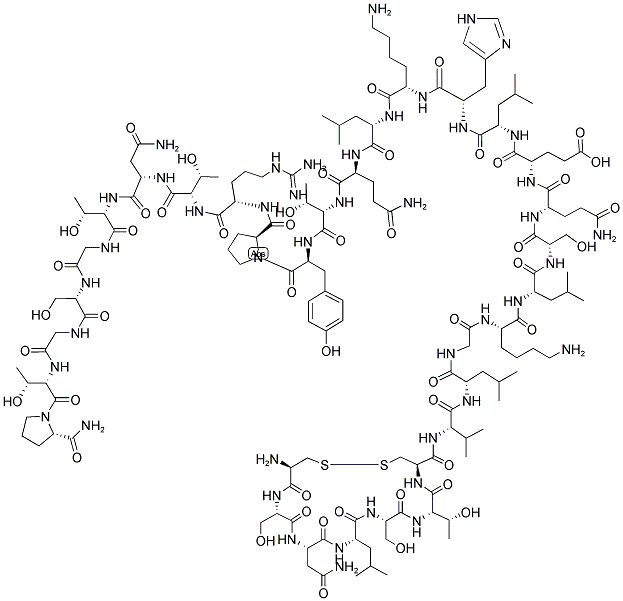
In vertebrates, a mature CT is composed of 32 aa residues. These CTs share a basic feature: a disulfide bridge between the cysteine residues at positions 1 and 7, which forms a ring of 7 aa residues at the N-terminal side. They also share a proline amide group at the C-terminal end. An invertebrate CT sequence was recently determined from the protochordate, Ciona intestinalis. The protochordate CT sequence was composed of 30 aa residues but was shown to display high similarity to that of vertebrate CTs. They share the N-terminal circular region and C-terminal amidated proline. Human CT has an α-helical structure between the ninth and 16th residues, and shows both hydrophilic and hydrophobic properties.
The mature CT sequences of nonmammalian vertebrates, including teleosts, urodeles, reptiles, and birds, are considerably conserved, although those of cartilaginous fish and anurans are slightly variant from those of other nonmammals. In mammals, the primary structure is quite different from that of nonmammals. Mammalian CTs share a conserved C-terminal amidated proline and an N-terminal circular structure, which is formed by a disulfide bridge between cysteine residues at positions 1 and 7.


Primary structure of a mature human CT.
Biological functions
Target cells/tissues and functions
Osteoclasts are the major target for the action of CT in mammals. As CT acts on the central nervous system (CNS), it prevents painful osteoporosis in humans. CT has specific binding sites within the CNS in mammals. The intracerebral injection of CT suppresses food and water intake in rats. In addition, CT functions in various tissues and organs, including the gastrointestinal tract, kidney, breast, and hypothalamo-pituitary axis.
Phenotype in gene-modified animals
αCGRP and CT are products of the same gene. αCGRP is predominantly produced in the nervous system through tissue-specific alternative splicing of the initial gene transcript. The human CT/αCGRP gene has six exons. The mRNA encoding the CT precursor consists of exons 1, 2, 3, and 4, and is predominantly expressed in thyroid C-cells. The mRNA encoding the αCGRP precursor in the nervous system contains exons 1, 2, 3, 5, and 6 . Ct/αCgrp-knockout mice experience increased bone mass and bone formation rate.11 Increased bone formation has also been observed in heterozygous Ctr+/- mice. Homozygous Ctr null mice cannot be studied because of the embryonic lethal before skeletal formation.
Pathophysiological implications
Clinical implications
CT reduces bone resorption by inhibiting mature active osteoclast formation, and increases renal calcium excretion. The activity of fish CTs is stronger than mammalian CTs. Therefore, fish CTs are used in the management of postmenopausal osteoporosis, Paget’s disease of the bone, Sudeck’s atrophy, and malignancy-associated hypercalcemia. In addition to its effect on osteoclasts and renal tubules, CT has an analgesic effect, possibly mediated through β-endorphins and the central modulation of pain perception. CT has been shown to relieve pain in patients with a number of conditions.
Use for diagnosis and treatment
The plasma CT concentration is usually measured using commercially available ELISA kits. The plasma CT level is a specific and sensitive marker of medullary thyroid carcinoma (MTC). Tumor size is significantly correlated with plasma CT levels, which can increase to more than 4000 pg/mL in MTC patients. ProCT is the most established biomarker used to diagnose and treat septic patients, and is available in clinical practice. ELISA kits for proCT and CT such as for humans, chickens, eels, and salmon are sold by various companies.