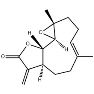
| Description | Parthenolide is an NF-κB inhibitor, reduces histone deacetylase 1 (HDAC-1) and DNA methyltransferase 1 independent of NF-κB inhibition. |
|---|
| Related Catalog | Signaling Pathways >> Autophagy >> Autophagy Signaling Pathways >> Autophagy >> Mitophagy Signaling Pathways >> NF-κB >> NF-κB Natural Products >> Terpenoids and Glycosides Research Areas >> Cancer |
|---|
| Target | NF-κB Autophagy Mitophagy |
|---|
| In Vitro | Parthenolide (PTL) has a dose-dependent growth inhibition effect on NSCLC cells Calu-1, H1792, A549, H1299, H157, and H460. Parthenolide can induce cleavage of apoptotic proteins such as CASP8, CASP9, CASP3 and PARP1 both in concentration- and time-dependent manner in tested lung cancer cells, indicating that apoptosis is trigged after Parthenolide exposure. In addition to induction of apoptosis, Parthenolide also induces G0/G1 cell cycle arrest in a concentration-dependent manner in A549 cells and G2/M cell cycle arrest in H1792 cells[2]. |
|---|
| In Vivo | Only Parthenolide, the HDAC inhibitor with anti-inflammatory features, displayed a potent anti-apoptotic effect in Phb1 KO hepatocytes. Indeed, TSA and Parthenolide-treated hepatocytes showed increased levels of FXR, and reduced levels of CYP7A1, HDAC4, TNFα, TRAIL and Bax suggesting a less toxic effect of bile acids as a results of specific HDAC inhibition, resulting in the attenuation of the Phb1 KO hepatocytes apoptotic response. Importantly, Parthenolide exerts a protective effect from the liver injury after BDL in Phb1 KO mice. Indeed, Parthenolide treatment results in a reduction of the mortality rate of this mice after BDL associated with a lower apoptotic response as revealed by a reduction of necrotic areas, Tunel-staining, as well as decreased ALT (8431±957 vs.4225±210 U/L) and AST (4805±300 vs.2242±438 U/L) activities compared to control Phb1 KO mice[3]. |
|---|
| Cell Assay | Human lung cancer cell lines are seeded in 96-well plates and treated on the second day with the given concentration of Parthenolide (0, 5, 10, 20 μM) for another 48 hours and then subjected to SRB or MTT assay. For SRB assay, live cell number is estimated as described earlier. After treatment, the medium is discarded firstly. In order to fix the adherent cells, 100 μL of cold trichloroacetic acid (10% (w/v)) are adding to each well and incubating at 4°C for at least 1 hour. The plates are then washed five times with deionized water and dried in the air. Each well are then added with 50 μL of SRB solution (0.4% w/v in 1% acetic acid) and incubated for 5 min at room temperature. The plates are washed five times with 1% acetic acid to remove unbound SRB and then air dried. The residual bound SRB is solubilized with 100 μL of 10 mM Tris base buffer (pH 10.5), and then read using a microtiter plate reader at 495 nm. The MTT assay is executed. 20 μL MTT (5 mg/mL) are added to each sample and incubate at 37°C for 4 h, then 100 μL solubilization solution are added. Cell viability is determined at 595 nm[2]. |
|---|
| Animal Admin | Mice[3] Phb1 KO mice are used. Males from 8-12 weeks of age are treated. Parthenolide is intraperitoneally injected at a dose of 3 mg/kg 24 h and 1h before bile duct ligation (BDL) or twice a week during two weeks. Liver specimens are snap-frozen for subsequent analysis[3]. |
|---|
| References | [1]. Nakshatri H, et al. NF-κB-dependent and -independent epigenetic modulation using the novel anti-cancer agent DMAPT. Cell Death Dis. 2015 Jan 22;6:e1608. [2]. Zhao X, et al. Parthenolide induces apoptosis via TNFRSF10B and PMAIP1 pathways in human lung cancer cells. J Exp Clin Cancer Res. 2014 Jan 6;33:3. [3]. Barbier-Torres L, et al. Histone deacetylase 4 promotes cholestatic liver injury in the absence of prohibitin-1. Hepatology. 2015 Oct;62(4):1237-48. |
|---|

 China
China