Melamine: Overview, Neurotoxicity and its Mechanism
General Description
Melamine, a nitrogen-rich chemical, is used primarily in the synthesis of melamine formaldehyde resins for the manufacture of laminates, plastics, coatings, commercial filters, glues or adhesives, and molded compounds such as dishware and kitchenware. When mixed with resins, melamine has fire retardant properties owing to its release of nitrogen gas when burned or charred. It is also used in paints and as a fertilizer. Low levels of melamine may also be present in water effluents as a result of industrial-scale uses, production, and disposal. It has received much attention in recent years owing to a series of highly publicized food safety incidents. These include pet food recalls in North America in 2007 and the deaths of six infants and the illness of some 300,000 more in China in 2008 owing to the adulteration of milk, infant formula, and other milk-derived products. The embryos and infants are in the developmental stages, and their organs are especially vulnerable, so the potential damages to the organs may be asymptomatic in a short time but last for a long time. This article will introduce the neurotoxicity of melamine and potential mechanism behind it.1
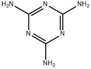
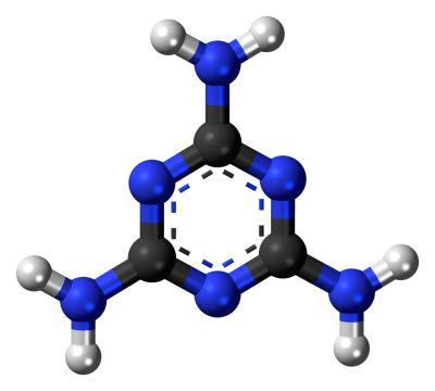
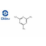
Figure 1. Melamine
Neurotoxicity
Despite that there is a common view that melamine has a half-life of approximately 3–4 h in animal studies, given the fact that infants are fed every 3 or 4 h and milk accounts for a large proportion in their diet, excessive melamine may deposit and cause damage to several organs. A selection of studies using neuronal cells indicated that melamine can induce apoptosis, disrupt metabolism, hyperpolarization, and spontaneous firing. MTT assay indicated that melamine inhibited the proliferation of differentiated cells in a concentration- and time-dependent manner. Hoechst 33258 staining and flow cytometry assay showed that 33 mg/ml melamine cultured for 24 h induced apoptotic cell death rather than necrosis. Wang et al. found that treatment with 312 mg/ml melamine stimulated for 12 h could induce pathologic changes in hippocampal neurons, such as shrinkage, dense chromatin, and insoluble metabolites. Besides, caspase-3 activity of hippocampal neurons after being challenged with melamine was higher than that of normal ones in the mass, which revealed that cells underwent apoptotic process.
Recent study showed that melamine enhanced autophagy through increasing ROS levels in mesangial cells. Their studies provided compelling evidence that low levels of melamine exposure may also represent a health risk to nerve. More importantly, the results indicated that neurons may be excessively vulnerable to melamine, which opposed the idea that damage of melamine was limited to the kidneys.2
Mechanism
There has been difficulty in development of a rationale concerning mechanisms underlying melamine neurotoxicity. Various suggestions have been made, including the possibility that melamine itself impaired hippocampal synaptic plasticity. This was supported by the observation that, gavaged with melamine, adolescent rats’ long-term potentiation (LTP) suppressed significantly through presynaptically and postsynatically inhibited glutamatergic transmission. Given these findings with LTP, the same study also examined the potential effects of melamine on long-term depression (LTD), while the fEPSP slope of LTD was enhanced markedly.
Recent study indicated that the NMDARs, but not AMPARs, were blocked by acute melamine and caused memory consolidation deficits. The activation of NMDA-2B subunits can rescue hippocampal LTD close to control level. In one study, investigating the synaptic NMDA receptor-mediated current, prenatal melamine exposure during brain growth spurt significantly decreased paired pulse ratio of the field excitatory postsynaptic currents. It demonstrated that the enhanced of PPF may be partly associated with the slightly initial probability of presynaptic neurotransmitter release in hippocampal CA1 neurons, being compatible with the suppressive synaptic efficiency in local field potential recordings. PPF function depended on presynaptic calcium dynamics. Previously, it reported that the significantly increased Ca2+ fluorescences of hippocampal neurons indicated the the toxicity of melamine to neurons was through disturbing the calcium homeostasis, which plays pivotal roles in diverse biological action in modulating and controlling neuronal excitability. In the mammalian brain, these two forms of plasticity were characterized by a long-lasting increase or decrease in synaptic strength, respectively.
Both processes were considered to be involved in information storage, so in learning, memory and other physiological processes. Moreover, the balance of synaptic transmission strongly supported the hypothesis that common mechanisms underlying the association synaptic plasticity balance with behavioral plasticity. Therefore, the behavioral consequence of their inhibition could be due to the impairment of LTP, LTD, or inhibition of synaptic transmission.2
1. A Brief Review of Neurotoxicity Induced by Melamine. DOI: 10.1007/s12640-017-9731-z
References:
[1] LEI AN W S. A Brief Review of Neurotoxicity Induced by Melamine.[J]. Neurotoxicity Research, 2017, 32 2. DOI:10.1007/s12640-017-9731-z.You may like
Related articles And Qustion
See also
Lastest Price from Melamine manufacturers
US $0.00/kg2025-11-19
- CAS:
- 108-78-1
- Min. Order:
- 1kg
- Purity:
- 99%
- Supply Ability:
- 10000KGS
US $10.00/KG2025-04-21
- CAS:
- 108-78-1
- Min. Order:
- 1KG
- Purity:
- 99%
- Supply Ability:
- 10 mt
