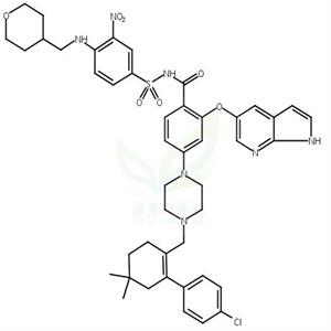
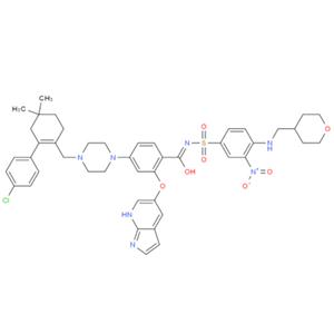
| 名称 | Venetoclax |
| 描述 | Venetoclax (ABT-199) is a Bcl-2 inhibitor (Ki<0.01 nM) with potent, selective, and orally active properties. Venetoclax has a 3-order-of-magnitude lower affinity for Bcl-xL and Bcl-W (Kis=48/245 nM). Venetoclax induces autophagy and apoptosis. |
| 细胞实验 | RS4;11 cells were seeded at 50,000 per well in 96-well plates and treated with compounds diluted in half-log steps starting at 1 μM and ending at 0.00005 μM. All other leukemia and lymphoma cell lines were seeded at 15,000–20,000 cells per well in the appropriate medium and incubated with ABT-199 or navitoclax for 48 h. Effects on proliferation were determined using Cell TiterGlo reagent. EC50 values were determined by nonlinear regression analysis of the concentration-response data. Mouse FL5.12–BCL-2 and FL5.12–BCL-XL cells were propagated and assessed as described previously. Bak?/? Bax?/? double knockout mouse embryonic fibroblasts were seeded into 96-well microtiter plates at 5,000 cells per well in DMEM supplemented with 10% FBS. ABT-199 in the same culture medium was added in half-log dilutions starting at 5 μM. The cells were then incubated at 37 °C (5% CO2) for 48 h, and the effects on proliferation were determined using Cell TiterGlo reagent according to the manufacturer's instructions [1]. |
| 激酶实验 | The equilibrium binding experiments of fluorescent peptides to Bcl-xL protein were performed in an Analyst 96-well plate reader under the following conditions: each individual well in a 96-well assay plate contained 5 μl DMSO, 15 nM fluorescent peptide, and increasing concentrations (from 0 to 2.24 μM) of Bcl-xL protein in assay buffer in a final volume of 125 μl. The plate was mixed on a shaker for 1 min and incubated at room temperature for an additional 15 min. The polarization in millipolarization units (mP) was measured at room temperature with an excitation wavelength at 485 nm and an emission wavelength at 530 nm. For assay stability testing, a plate containing a binding experiment was measured at different times over a 24-h period. Between each reading, the plate was covered with parafilm to prevent any solution evaporation. To determine the effect of DMSO on the assay, binding experiments were performed under conditions similar to those described above except that the amount of DMSO was varied from 0 to 4 to 8%. All experimental data were analyzed using Prism 3.0 software and Kd values were generated by fitting the experimental data using a sigmoidal dose-response nonlinear regression model [1]. |
| 动物实验 | Female C.B-17 SCID mice (DoHH2 and Granta-519 xenografts) and female C.B-17 SCID-beige mice (RS4;11 and Toledo xenografts) were inoculated with 1 × 10^6 (DoHH2) or 5 × 10^6 (Granta-519, Toledo and RS4;11) cells subcutaneously in the right flank. The inoculation volume (0.2 ml) comprised a 50:50 mixture of cells in growth media and Matrigel. Electronic calipers were used to measure the length and width of each tumor 2–3 times per week. Tumor volume was estimated by applying the following equation: volume = length × width2/2. When tumors reached approximately 220 mm3, mice were size matched (day 0) into treatment and control groups. All xenograft trials were conducted using ten mice per group, and all mice were ear tagged and monitored individually throughout the studies. ABT-199 was formulated for oral dosing in 60% phosal 50 propylene glycol (PG), 30% polyethylene glycol (PEG) 400 and 10% ethanol, and bendamustine and rituximab were formulated in accordance with the manufacturer's instructions. ABT-199 was delivered approximately 2 h before bendamustine or bendamustine plus rituximab. TGImax was calculated as the greatest treatment response using the following equation: TGImax = (1 ? mean tumor volume of the treated group/mean tumor volume of the vehicle control group) × 100. The TGD (%) was determined as the percentage increase of the median time period for the treatment group to reach an arbitrary tumor volume of 1,000 mm3 relative to the vehicle control group. A complete tumor regression response was the portion of the population with tumors ≤25 mm3 for at least three consecutive measurements [1]. |
| 体外活性 | 方法:11 种人 T 细胞急性淋巴细胞白血病细胞 T-ALL 用 Venetoclax (0-12 μM) 处理 48 h,使用 Celltiter-Glo Luminescent Cell Viability Assay 检测细胞活力。
结果:Venetoclax 对 11 种 T-ALL 的 IC50 值范围为 0.2-10 μM。[1]
方法:人急性淋巴白血病细胞 RS4;11 用 Venetoclax (0.01-5 μM) 孵育 3.5 h,使用 Caspase-GLO kit 评估 Caspase-3/7 活性。
结果: Venetoclax 诱导 Caspase 的激活,这是细胞凋亡特征之一。[2]
方法:人原代 HCL 白血病细胞用 Venetoclax (0.1-1 μM) 处理 24 h,使用 Flow Cytometry 方法检测细胞死亡情况。
结果:Venetoclax 以剂量依赖的方式显著增加了 HCL 细胞的细胞死亡。[3] |
| 体内活性 | 方法:为检测体内抗肿瘤活性,将 Venetoclax (100 mg/kg in 60% PG+30% PEG 400+10% ethanol) 口服给药给携带人弥漫大B细胞淋巴瘤 Toledo 的 C.B-17 SCID-beige 小鼠,每天一次,持续二十一天。
结果:Venetoclax 可显著抑制 Toledo 肿瘤的生长 (TGImax=93%,TGD=220%)。[2]
方法:为检测体内抗肿瘤活性,将 Venetoclax (50 mg/kg in 10% ethanol+30% PEG 400+60% Phosal 50PG,口服给药,每天一次) 和 anti-PD-1 (10 mg/kg in PBS,腹腔注射,每四天三次) 给药给携带小鼠结直肠癌肿瘤 MC38 的 C57BL/6 小鼠,持续十四天。
结果:Venetoclax 可以增强免疫检查点抑制剂 (ICIs) 的抗肿瘤功效,同时增加 PD-1+T 效应记忆细胞。Venetoclax 在体外对抗原刺激的反应中不会损害人类 T 细胞的功能,也不会拮抗 anti-PD-1 诱导的 T 细胞活化。[4] |
| 存储条件 | Powder: -20°C for 3 years | In solvent: -80°C for 1 year | Shipping with blue ice. |
| 溶解度 | H2O : < 1 mg/mL (insoluble or slightly soluble)
Ethanol : < 1 mg/mL (insoluble or slightly soluble)
DMSO : 100 mg/mL (115.15 mM)
|
| 关键字 | bioavailable | GDC0199 | orally | inhibit | Inhibitor | Bcl-2 Family | GDC 0199 | Venetoclax | Autophagy |
| 相关产品 | Oxyresveratrol | Guanidine hydrochloride | Naringin | Taurine | Gefitinib | Hydroxychloroquine | Curcumin | Stavudine | Paeonol | Sodium 4-phenylbutyrate |
| 相关库 | 抑制剂库 | 抗癌活性化合物库 | 抗癌上市药物库 | 已知活性化合物库 | EMA 上市药物库 | 高选择性抑制剂库 | FDA 上市药物库 | 药物功能重定位化合物库 | 抗癌临床化合物库 | 抗癌药物库 |