
7YR企业会员
发布人:爱必信(上海)生物科技有限公司
发布日期:2026/7/3 14:20:06
在结直肠癌(CRC)治疗中,肿瘤相关成纤维细胞(CAFs)引发的免疫抑制微环境是免疫治疗响应率低的核心症结。近期发表于《Nature Communications》的一项突破性研究(PMID: 41690961),提出了基于 Cu@Fe₃O₄磁性纳米颗粒(NPs)的铁死亡纳米疗法,通过精准靶向 CAFs 重塑肿瘤微环境,为 CRC 治疗开辟了新路径。爱必信(Absin)的 α-SMA 抗体(货号:abs172181)作为关键工具,全程助力 CAFs 的鉴定与功能验证,为研究的顺利推进提供了可靠支撑。
文献标题:Amelioration of colorectal cancer-associated fibroblasts in immunosuppressive microenvironment by ferroptosis-based nanotherapy
发表期刊:Nat Commun. (IF=15.7)
DOI:https://doi.org/10.1038/s41467-026-69462-5
使用 Absin 产品:Rabbit anti-α Smooth Muscle Actin Recombinant Monoclonal Antibody(AF647 Conjugate) (132-63)(货号:abs172181)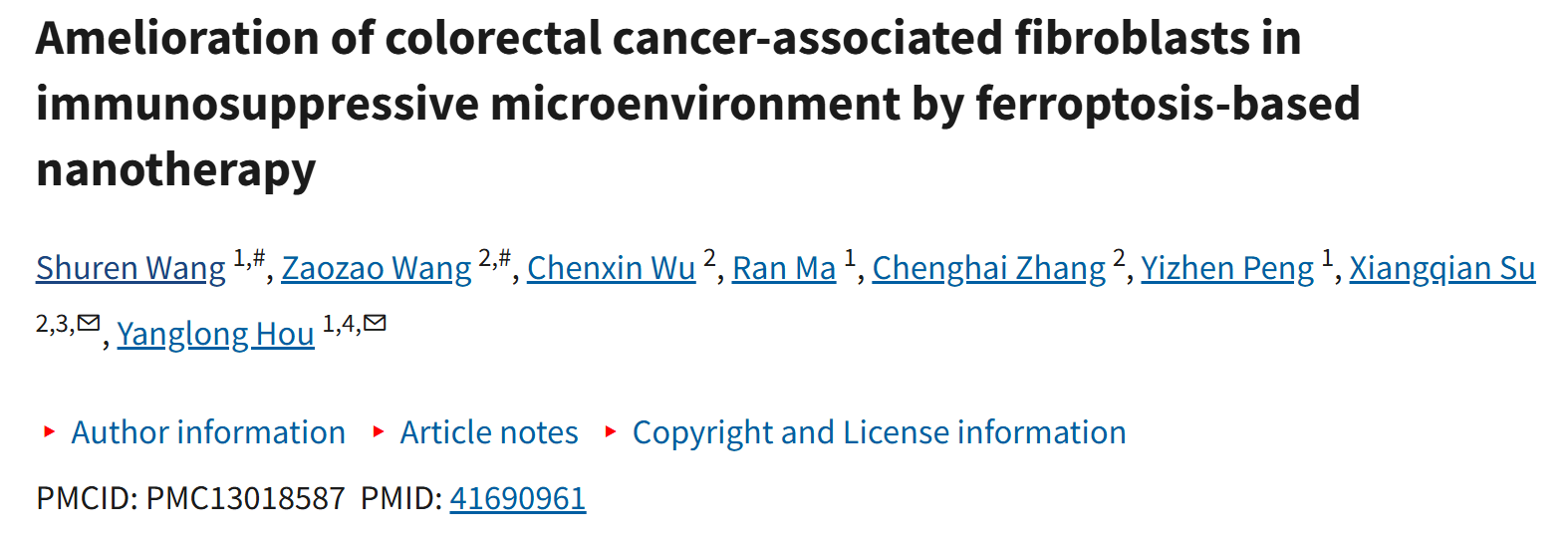

85% 的微卫星稳定型 CRC 对免疫治疗不敏感,根源在于 CAFs 的双重破坏作用:一方面通过分泌细胞外基质(ECM)形成物理屏障,阻碍药物渗透与 T 细胞浸润;另一方面通过分泌 TGF-β 等细胞因子诱导免疫抑制,导致 T 细胞耗竭、树突状细胞(DCs)功能异常。传统靶向 CAFs 策略存在异质性强、副作用大等瓶颈,亟需开发高效低毒的新型疗法。
研究团队跳出传统 "直接杀伤肿瘤细胞" 的思维定式,提出 "靶向 CAFs 重塑免疫微环境" 的治疗框架:
设计核心 - 壳结构的 Cu@Fe₃O₄ NPs,利用铁离子介导的活性氧(ROS)生成和铜离子介导的谷胱甘肽过氧化物酶 4(GPX4)下调,协同诱导 CAFs 铁死亡;
借助 NPs 的光热转换特性与过氧化物酶样活性,实现光热治疗(PTT)与化学动力学治疗(CDT)的协同增效;
通过调节 CAFs 趋化因子分泌(上调 CCL3、下调 CXCL12),促进 DCs 成熟与 CD8⁺T 细胞活化,将免疫抑制微环境转化为免疫激活状态;
结合 AS1411 适配体靶向 CAFs 高表达的核仁素(NCL),提升肿瘤靶向特异性(图 1a)。
Fig. 1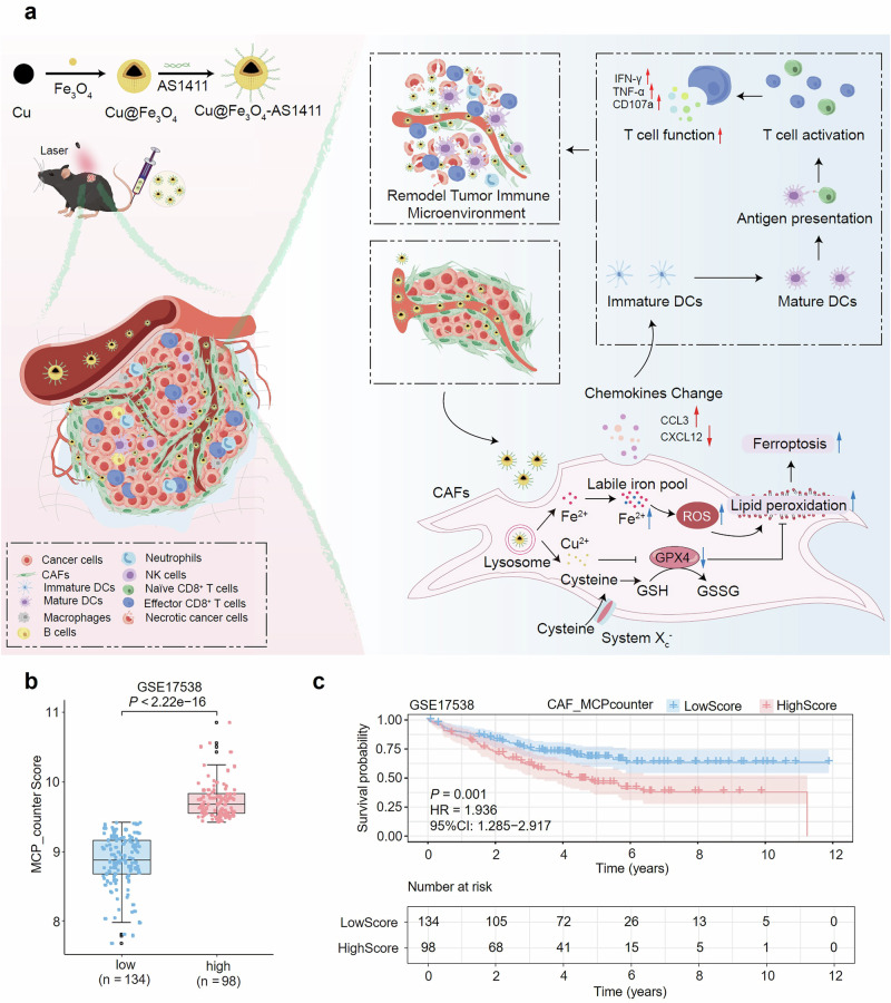
a Schematic illustration of the antitumor process of NPs through inducing the ferroptosis of CAFs and remodeling the CAFs-related immune TME. b Individuals in the GSE17538 cohort are stratified into low and high CAF groups based on MCP-counter derived CAF abundance by the optimal cutoff value. Box plots depict the median (centre line), interquartile range (25th-75th percentiles; box), and 1.5 × interquartile range (whiskers). For the low group: min = 7.682, Q1 = 8.675, median = 8.873, Q3 = 9.165, max = 9.414 (n = 134 patients); for the high group: min = 9.424, Q1 = 9.547, median = 9.673, Q3 = 9.826, max = 10.854 (n = 98 patients). Statistical significance is assessed using a two-sided Mann-Whitney U test. c Kaplan-Meier curves of 232 colon cancer patients from the GSE17538 cohort stratified by CAF abundance assessed using the MCP-counter method. Tick marks indicate censored events. P values are calculated using a two-sided log-rank test, and hazard ratios (HRs) with 95% confidence intervals are estimated using univariate Cox proportional hazards regression. Source data are provided as a Source Data file.
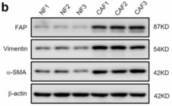
研究首先从 CRC 患者组织中成功分离 CAFs 与正常成纤维细胞(NFs),通过 Western blot 验证 CAFs 高表达 FAP、Vimentin 及 α-SMA 等特征标志物(图 2b)。体外实验证实,Cu@Fe₃O₄ NPs 对 CAFs 的细胞毒性显著高于 NFs(图 2c),且在 808nm 激光照射下,通过 ROS 爆发与 GPX4 下调的协同作用,实现 CAFs 的高效铁死亡(图 2e-g)。
Fig. 2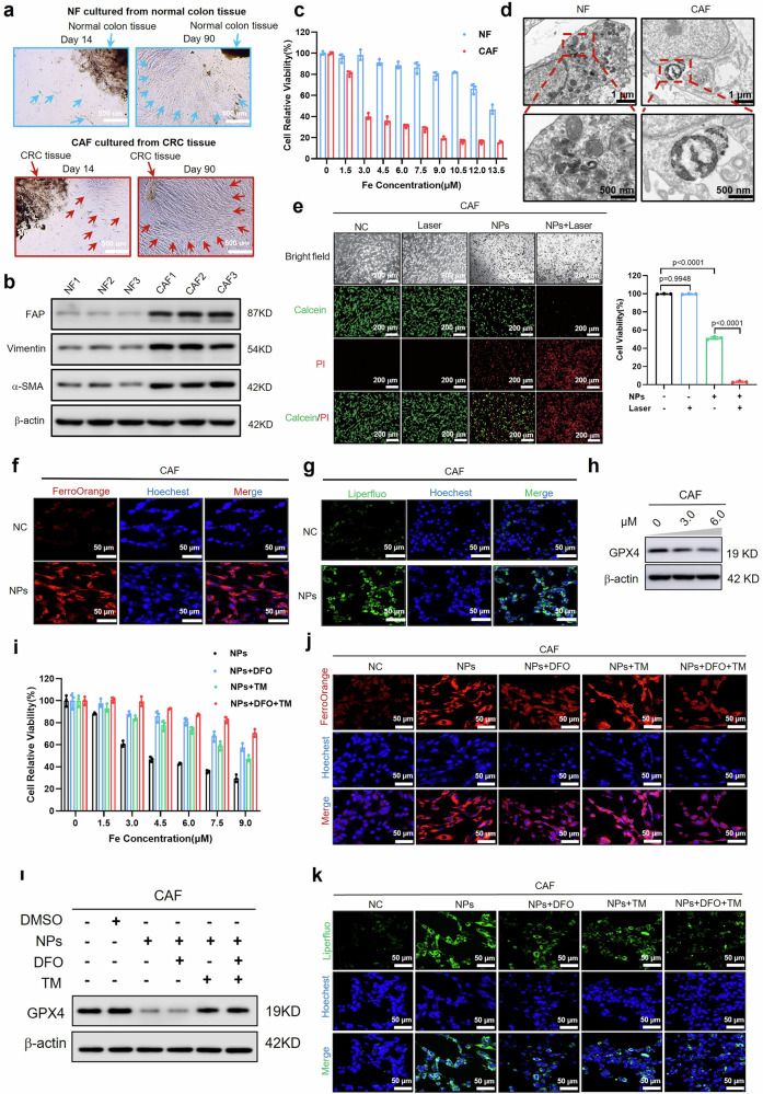
a Images displaying the isolation of NFs from adjacent normal tissues and CAFs from CRC tumor tissues. b Western blot confirming successful isolation of NFs from three normal colonic tissues and CAFs from three CRC specimens, validated by antibodies against fibroblast activation protein-α (FAP), Vimentin, and α-smooth muscle actin (SMA); β-actin as a loading control. c Cytotoxicity assessment of NPs in NFs and CAFs for 24 h at varying concentrations. d Bio-TEM images of NFs and CAFs incubated with NPs for 24 h; Red dashed boxes marking NPs location. e Bright-field and fluorescent images of CAFs stained with Calcein-AM (live cells, green fluorescence) and PI (dead cells, red fluorescence) after treatment with phosphate buffer saline (PBS), Laser, NPs, and NPs+Laser. PBS as control (NC) (left); Statistical analysis of cell viability (right). f, g Fluorescent images of (f) intracellular Fe2+ levels and (g) LPO in CAFs treated without or with NPs for 24 h; Nuclei stained with Hoechst 33342 (blue). h Western blot of GPX4 expression in CAFs treated with increasing concentrations of NPs for 24 h; β-actin as a loading control. i Relative cell viability of CAFs treated with NPs alone, NPs with DFO, NPs with TM, and NPs with both DFO and TM for 24 h by CCK8 assay. j, k Fluorescent images of (j) intracellular Fe2+ levels and (k) LPO in CAFs treated as in (i); Nuclei stained with Hoechst 33342 (blue). l Western blot of GPX4 expression in CAFs treated as in (i); β-actin as a loading control. n = 3 independent experiments per group in (c–g) and (i–k). Western blot experiments in (b, h, l), FerroOrange and Liperfluo staining experiments in (f, g, j, k) are repeated independently three times with similar results. Data are presented as means ± standard deviation (SD). Statistical analyses are performed using one-way analysis of variance (ANOVA) with multiple comparisons (e). Source data are provided as a Source Data file.
NPs 处理后的 CAFs 分泌谱发生显著改变:促炎趋化因子 CCL3 表达上调,免疫抑制趋化因子 CXCL12 表达下调(图 3k-l)。这一变化直接促进 DCs 表面共刺激分子 CD80/CD86 的表达(图 4g),并增强 CD8⁺T 细胞的活化标志物(T-bet/Eomes)与功能分子(IFN-γ、TNF-α、CD107a)表达(图 4h-i),有效逆转了 CAFs 介导的免疫抑制。
Fig. 3
a Schematic illustration of cell viability and transwell migration assays using HCT116 cells treated with supernatants from NFs, CAFs, or NPs-treated CAFs. b Transwell migration images presenting the number of migrated HCT116 cells treated with CM from NFs, CAFs, or NPs-treated CAFs for 24 h. c Cell viability of HCT116 cells treated as in (b) by the CCK8 assay. d Western blot of E-cadherin, N-cadherin, and Vimentin expression in HCT116 cells treated with CM from NFs, CAFs, or NPs-treated CAFs for 24 h; β-actin as a loading control. e, f Bubble plots of (e) GO and (f) KEGG pathway enrichment analyses for differentially expressed genes (DEGs) in NPs-treated CAFs compared to controls. g, h MSigDB Hallmark GSEA analysis showing enrichment of (g) chemokine activity and (h) ferroptosis pathways in NPs-treated CAFs compared to controls. ES: enrichment score; NES: normalized enrichment score. i Heatmap and histogram displaying gene expression levels of DEGs (log₂|FC | ≥ 1) from three chemokine-related GO pathways (GO:0016493, GO:0048020, GO:0008009) in NPs-treated CAFs compared to controls, ranked by differential expression. j QRT-PCR quantification of CCL3, CXCL12, and CCR7 expression in NPs-treated CAFs compared to controls. k, l Anti-human ELISA quantification of (k) CCL3 and (l) CXCL12 concentrations in supernatants from CAFs treated without or with NPs for 24 h. m Western blot of MAPK, p-MAPK (Thr180/Tyr182), NF-κB, p-NF-κB (Ser536), AKT, and p-AKT (Ser473) expression in CAFs treated without or with NPs for 24 h; β-actin as a loading control. n = 3 independent experiments in (b, c) and (j–l). Western blot experiments in (d, m) are repeated independently three times with similar results. Data are presented as means ± SD. Statistical analyses are performed using one-way ANOVA with multiple comparisons for (b, c); and two-tailed unpaired Student's t-test for (j–l). Source data are provided as a Source Data file.
Fig. 4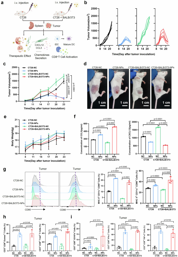
a Schematic illustration of the treatment strategy and related immune assessment in vivo based on NPs for the CT26 and the CT26 + BALB/3T3 group. b Individual tumor growth curves and (c) average tumor growth curves of the CT26 and the CT26 + BALB/3T3 group without or with the treatment of NPs. d Representative images of mice bearing CT26 cells and CT26 + BALB/3T3 cells without or with the treatment of NPs. e Body weight curves of CT26 cells- and CT26 + BALB/3T3 cells-bearing mice without or with the treatment of NPs. f Quantified expression of CCL3 and CXCL12 in homogenized tumor tissues from the CT26 and the CT26 + BALB/3T3 groups without or with the treatment of NPs. g Flow cytometry and corresponding statistical analysis of CD11c+MHC II+CD80+ and CD11c+MHC II+CD86+ cells (in total CD45+ cells) around tumor tissues of mice in four groups. h Statistical analysis of flow cytometry of CD3+CD8+Eomes+ and CD3+CD8+T-bet+ cells around tumor tissues of mice in four groups. i Statistical analysis of flow cytometry of CD3+CD8+CD107a+, CD3+CD8+IFN-γ+, and CD3+CD8+TNF-α+ cells around tumor tissues of mice in four groups. n = 5 mice per group in (b–e) and n = 3 independent experiments per group in (f–i). Data are presented as means ± SD. Statistical analyses are performed using two-way ANOVA with multiple comparisons for (c); and one-way ANOVA with multiple comparisons for (f–i). Source data are provided as a Source Data file.
在皮下肿瘤模型、基因工程小鼠模型(GEMM)、患者来源异种移植模型(PDX)及类器官模型(PDO)中,NPs + 激光治疗均展现出显著的肿瘤抑制效果。其中 PDX 模型中,NPs-AS1411 + 激光组肿瘤几乎完全消退(图 7b-c),且对正常组织无明显损伤,证实了疗法的安全性与临床转化潜力。
Fig. 7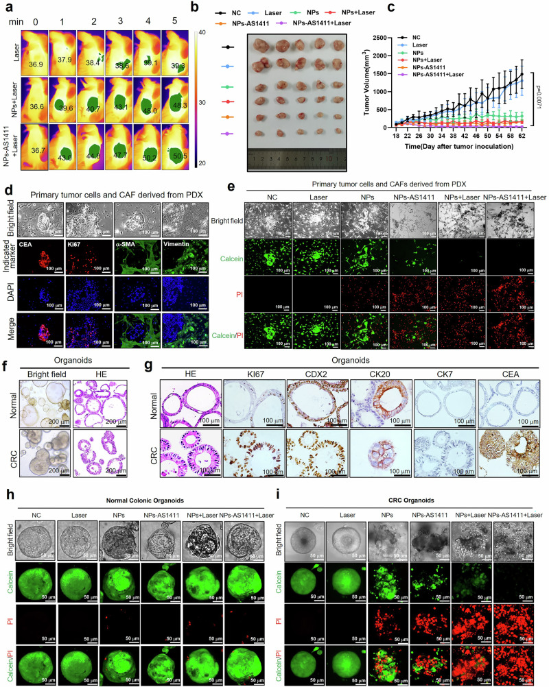
a Real-time thermal infrared images of the PDX model after intravenous injection of PBS, NPs, and NPs-AS1411 for 24 h under 808 nm laser irradiation. b Representative images of tumors of the PDX model in six groups. c Average tumor growth curves of the PDX model in six groups. d Bright-field and fluorescent images of CEA, Ki67, α-SMA, and Vimentin expression in co-cultured tumor cells and CAFs from PDX model tumor tissues. e Bright-field and fluorescent images of co-cultured tumor cells and CAFs from PDX model tumor tissues stained with Calcein-AM (live cells, green fluorescence) and PI (dead cells, red fluorescence) after treatment with PBS, Laser, NPs, NPs-AS1411, NPs + Laser, and NPs-AS1411 + Laser. f Bright-field and H&E staining images of organoids from normal colonic epithelia and CRC. g H&E staining and immunohistochemical images of expression of Ki67, CDX2, CK20, CK7, and CEA of normal colonic organoids and CRC organoids. h, i Confocal bright-field and fluorescent images of Calcein-AM (live cells, green fluorescence) and PI (dead cells, red fluorescence) staining in (h) normal colonic organoids and (i) CRC organoids after treatment with PBS, Laser, NPs, NPs-AS1411, NPs + Laser, and NPs-AS1411 + Laser. n = 5 mice per group in (a–c). Immunofluorescent experiments (d, e), H&E and immunohistochemical experiments (f, g), and Calcein-AM/PI staining experiments (h, i) are repeated three times with similar results. Data are presented as means ± SD. Statistical analyses are performed using two-way ANOVA with multiple comparisons for (c). Source data are provided as a Source Data file.
在整个研究中,爱必信的 α-SMA 抗体(abs172181)贯穿 CAFs 的分离鉴定、功能验证与体内定位全过程,成为不可或缺的实验支撑:
研究从 CRC 患者肿瘤组织中分离原代成纤维细胞后,利用 abs172181 抗体通过 Western blot 检测(图 2b)与免疫荧光染色(图 7d),验证了 CAFs 高表达 α-SMA 的特征表型,确保了实验材料的纯度与可靠性,为后续铁死亡诱导及信号通路分析奠定基础。

在 NPs 处理后的功能验证中,通过 abs172181 抗体检测发现,NPs 不仅诱导 CAFs 铁死亡,还显著下调其 α-SMA 表达,表明 CAFs 的活化表型被有效抑制,减少了 ECM 分泌与免疫抑制因子释放,直接证实了疗法对 CAFs 功能的重塑作用。
在体内实验中,abs172181 抗体通过免疫组化染色,清晰呈现了 CAFs 在肿瘤组织中的分布特征,以及 NPs 治疗后 CAFs 数量的显著减少(图 7d),为纳米颗粒的靶向富集效果与治疗机制提供了直接的组织学证据。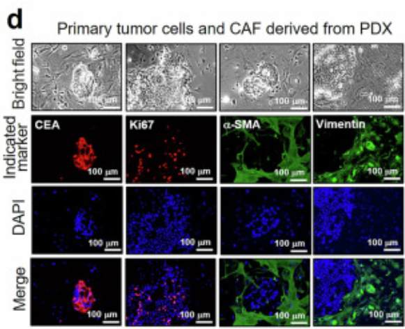
作为成纤维细胞活化的经典标志物,α-SMA 的精准检测是 CAFs 相关研究的核心技术节点。爱必信 abs172181 抗体凭借高特异性与高灵敏度,在 Western blot、免疫荧光、免疫组化等多种实验场景中表现稳定,完美匹配了本研究从体外细胞实验到体内动物模型的全方位验证需求,成为科研成果可靠性的重要保障。
该研究首次报道了 Cu@Fe₃O₄ NPs 通过铁死亡途径靶向 CAFs 的结直肠癌治疗策略,实现了 "物理屏障破除 + 免疫激活" 的双重目标,其创新点在于将纳米材料的理化特性与 CAFs 的生物学特征深度结合,为解决免疫治疗耐药提供了全新思路。
爱必信始终致力于为生命科学研究提供高品质工具试剂,除 α-SMA 抗体(abs172181)外,还提供 FAP、Vimentin 等 CAFs 相关标志物抗体,以及铁死亡、免疫细胞功能验证等系列产品,全方位覆盖肿瘤微环境研究的核心需求。未来,随着纳米疗法向临床转化,这类精准靶向肿瘤微环境的策略有望成为结直肠癌及其他实体瘤的新一代治疗方案,而爱必信也将持续为科研与临床转化搭建桥梁,助力更多突破性研究的诞生。
如需了解 abs172181 抗体的详细实验方案或相关产品组合,可访问爱必信官网或联系技术支持团队,获取定制化解决方案。
相关新闻资讯