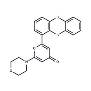
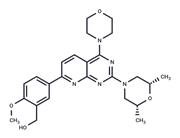
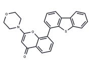
| Name | KU-55933 |
| Description | KU-55933 (ATM Kinase Inhibitor) is an ATM inhibitor (IC50=12.9 nM; Ki=2.2 nM) that is selective and ATP-competitive. KU-55933 induces apoptosis and has antitumor effects. |
| Cell Research | U2OS cells are exposed to ionizing radiation (3, 5, or 15 Gy) or UV (5 or 50 J/m2) and the ATM response determined by Western blot analysis of p53 serine 15 phosphorylation and stabilization of wild-type p53. Whole cell extracts are obtained from each time point, proteins separated by SDS-PAGE, and the ATM-specific increase in phosphorylated serine 15 measured with a p53 phospho-serine 15 specific antibody. Overall p53 stabilization with time is also observed with a p53-specific antibody (DO-1). Similarly, for studying ATM-dependent phosphorylations on H2AX, CHK1, NBS1, and SMC1, the following antibodies are used: CHK1 phospho-serine 345 and NBS1 phospho-serine 343 antibodies. Histone H2A (H-124) and CHK1 antibodies are also used, as well as SMC1 and SMC1 phospho-serine 966 antibodies. For determination of a cellular IC50 for KU-55933, the peak response time for p53 serine 15 phosphorylation of 2 hours is used to monitor inhibition of ATM. KU-55933 is titrated onto cells and preincubated for 1 hour before ionizing radiation. Using scanning densitometry, the percentage inhibition relative to vehicle control is calculated, and the IC50 value is calculated as for the in vitro determinations.(Only for Reference) |
| Kinase Assay | Purified enzyme assays: ATM for use in the in vitro assay is obtained from HeLa nuclear extract by immunoprecipitation with rabbit polyclonal antiserum raised to the COOH-terminal 400 amino acids of ATM in buffer containing 25 mM HEPES (pH 7.4), 2 mM MgCl2, 250 mM KCl, 500 μM EDTA, 100 μM Na3VO4, 10% v/v glycerol, and 0.1% v/v Igepal. ATM-antibody complexes are isolated from nuclear extract by incubating with protein A-Sepharose beads for 1 hour and then through centrifugation to recover the beads. In the well of a 96-well plate, ATM-containing Sepharose beads are incubated with 1 μg of substrate glutathione S-transferase–p53N66 (NH2-terminal 66 amino acids of p53 fused to glutathione S-transferase) in ATM assay buffer [25 mM HEPES (pH 7.4), 75 mM NaCl, 3 mM MgCl2, 2 mM MnCl2, 50 μM Na3VO4, 500 μM DTT, and 5% v/v glycerol] at 37 °C in the presence or absence of inhibitor. After 10 minutes with gentle shaking, ATP is added to a final concentration of 50 μM and the reaction continued at 37 °C for an additional 1 hour. The plate is centrifuged at 250 × g for 10 minutes (4 °C) to remove the ATM-containing beads, and the supernatant is removed and transferred to a white opaque 96-well plate and incubated at room temperature for 1.5 hours to allow glutathione S-transferase-p53N66 binding. This plate is then washed with PBS, blotted dry, and analyzed by a standard ELISA technique with a phospho-serine 15 p53 antibody. The detection of phosphorylated glutathione S-transferase-p53N66 substrate is performed in combination with a goat antimouse horseradish peroxidase-conjugated secondary antibody. Enhanced chemiluminescence solution is used to produce a signal and chemiluminescent detection is carried out. |
| In vitro | METHODS: Ovarian cancer cells SKOV-3 were treated with Eflornithine hydrochloride hydrate (1-100 µM) for 48 h. Cell viability was measured by PrestoBlue assay.
RESULTS: Eflornithine significantly inhibited the viability of SKOV-3 cells in a dose-dependent manner. [1]
METHODS: MYCN2 (-) and MYCN2 (+) NB cells were treated with Eflornithine hydrochloride hydrate (5 mM) and putrescine/spermidine/spermine for 72 h. Cell migration was detected by wound healing assay.
RESULTS: Significant differences in cell migration were observed between untreated control cells and Eflornithine-treated cells. eflornithine inhibited cell migration by 73% and 72% in MYCN2 (-) and MYCN2 (+) cells, respectively. Supplementation of the medium with polyamines attenuated the effect of Eflornithine on MYCN2 (-) and MYCN2 (+) cells. [2] |
| In vivo | METHODS: To test the anti-infective activity in vivo, Eflornithine hydrochloride hydrate (25-50 mg/kg) and hydroxyurea (50-100 mg/kg) were orally administered to B. microti infected BALB/c mice once daily for 5 days.
RESULTS: Oral administration of hydroxyurea and Eflornithine inhibited the proliferation of Bifidobacterium microti in mice by 60.1% and 78.2%, respectively. hydroxyurea-DA and Eflornithine-DA combination treatment showed higher efficacy of chemotherapy than monotherapy. [3] |
| Storage | Powder: -20°C for 3 years | In solvent: -80°C for 1 year | Shipping with blue ice. |
| Solubility Information | Ethanol : 19.8 mg/mL (50 mM)
DMSO : 16.67 mg/mL (42.14 mM), Sonication is recommended.
|
| Keywords | KU55933 | Inhibitor | inhibit | Ataxia telangiectasia mutated | KU 55933 | ATM/ATR | ATM and RAD3 related | KU-55933 | Autophagy |
| Inhibitors Related | Stavudine | Xylitol | Myricetin | Sodium 4-phenylbutyrate | Hydroxychloroquine | Guanidine hydrochloride | Taurine | Curcumin | Oxyresveratrol | Paeonol | Naringin | Gefitinib |
| Related Compound Libraries | Highly Selective Inhibitor Library | Glycolysis Compound Library | Bioactive Compound Library | Tyrosine Kinase Inhibitor Library | Kinase Inhibitor Library | Inhibitor Library | Anti-Aging Compound Library | Bioactive Compounds Library Max | Anti-Cancer Active Compound Library | Neuronal Differentiation Compound Library |

 United States


