Derived from the root of Dipsacus asperoides CY Cheng et T .M. Ai.
Asperosaponin VI is a steroidal extract with anti-inflammatory and antinociceptive activity. long term use has been seen to attenuate cardiac dysfunction.
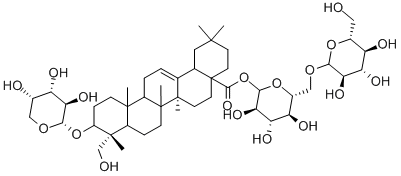
ChEBI: Akebia saponin D is a triterpenoid saponin that is hederagenin attached to a alpha-L-arabinopyranosyl residue at position 3 via a glycosidic linkage and a 6-O-beta-D-glucopyranosyl-beta-D-glucopyranosyl residue at position 28 via an ester linkage. It is the active ingredient found in the traditional Chinese herb Radix Dipsaci. It has a role as an apoptosis inducer, an antineoplastic agent, a plant metabolite, an anti-inflammatory agent, a bone density conservation agent, a neuroprotective agent and an antilipemic drug. It is a triterpenoid saponin, a trisaccharide derivative, a pentacyclic triterpenoid and a carboxylic ester. It is functionally related to a hederagenin.
Akebia saponin D (ASD) is a bioactive triterpenoid saponin isolated from the rhizome of Dipsacus asper Wall th at is used as an anti-osteoporosis drug. Akebia saponin D exhibits therapeutic effects in number of disease models including cancer, Alzheimerμs disease, cardiovascular disease, and bone fractures. Akebia saponin D protects against nonalcoholic fatty liver disease (NAFLD) liver damage in mice model of NAFLD. ASD decreases hepatic steatosis and heptocyte apoptosis through autophagy modulation. Akebia saponin D prevents oleic acid induced lipid droplets accumulation and increases autophagic flux BRL cells.
Asperosaponin VI (20 mg/kg; intravenous injection; 3 weeks) can accelerate wound healing and angiogenesis in a full-thickness cutaneous wound model of rats[3].
Asperosaponin VI (40 mg/kg; intraperitoneal injection; 3 weeks) has antidepressant effects in mice with chronic mild stress[4].
| Animal Model: | Male 8-week-old C57BL/6 mice treated chronic mild stress (CMS)[4] |
| Dosage: | 40 mg/kg |
| Administration: | Intraperitoneal injection (i.p.); 3 weeks |
| Result: | Significantly improved depression-like behaviors in CMS-induced mice, as shown by increased sucrose preference, prolonged latency in the forced swimming test and feeding latency in the novelty-suppressed feeding test, and shortened immobility time in the tail suspension test and forced swimming test.
Partially reversed CMS-induced weight loss.
Induced hippocampal microglia to switch from a pro-inflammatory to a neuroprotective phenotype, and regulated the levels of pro-inflammatory and anti-inflammatory cytokines.
Exerted anti-inflammatory and antidepressant effects through PPAR-γ pathway.
Promoted microglia-neuron crosstalk and prevented CMS-induced synaptic dysfunction of hippocampal neurons.
|