Dodecyl Sodium Sulfate
- Product NameDodecyl Sodium Sulfate
- CAS751-21-3
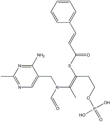
- MFC21H25N4O6PS
- MW492.49
- EINECS205-788-1
- MOL File751-21-3.mol
Chemical Properties
| Melting point | 204-207 °C(lit.) |
| Boiling point | 776.8±70.0 °C(Predicted) |
| Density | 1.03 g/mL at 20 °C |
| solubility | H2O: 0.1 M, clear to nearly clear, colorless to slightly yellow |
| form | Powder |
| pka | 1.84±0.10(Predicted) |
| color | White to slightly beige |
| CAS DataBase Reference | 751-21-3(CAS DataBase Reference) |
Safety Information
| Hazard Codes | Xi |
| Risk Statements | 36/37/38 |
| Safety Statements | 26-36/37 |
| RIDADR | UN 2926 4.1/PG 2 |
| WGK Germany | 2 |
| RTECS | WT1050000 |
| F | 3 |
MSDS
| Provider | Language |
|---|---|
| ACROS | English |
| SigmaAldrich | English |
| ALFA | English |
Usage And Synthesis
Sodium Lauryl Sulfate (SDS) is one of the most commonly used detergent in house holds and in Industry. It is a component of a number of industrially useful products. After use, like all other xenobiotics, it is discharged in water bodies in huge amounts.
Sodium Dodecyl Sulfate (SDS), a primary alkyl sulfate is a member of Alcohol sulfate family. Synthetic primary alkyl sulfates are based on feedstock derived from long-chain olefins by the use of the oxo process, which yields a mixture of linear and branched primary alcohols. Sulfonation of the mixed alcohols produces a mixture of linear primary alkyl sulfates (LPAS) and branched primary alkyl sulfates (BPAS), which have excellent detergent properties and are widely used in heavy-duty detergent applications. SDS denoted by molecular formula NaC12H25SO4, has a molecular weight of 288.38 g mol−1. The SDS molecule combines a nonpolar hydrophobic region with a strongly polar anionic end-group, thus mimicking, and competing with, certain membrane lipids. The peculiar structure of SDS renders this small amphipathic molecule (MW 288.4) highly suitable for complex formation both with nonpolar side chains and charged groups of amino acid residues in polypeptides of all possible sizes and shapes, without rupturing covalent bonds.

Figure 1 the chemical structure of SDS SDS synthesis is a relatively simple process involving the sulfation of 1-dodecanol followed by neutralization with a cation source. Purification is accomplished through repeated extraction. It is available commercially in both broad-cut and purified forms[1].
SDS is widely used in household products such as, toothpaste’s, shampoos, shaving foams, bubble baths, and cosmetics. In industry it is used as leather softening agent, wool cleaning agent, in paper industry as penetrant, flocculating agent, de-inking agent, in building construction as additive of concrete, oil well fire fighting, fire fighting devices, engine degreasers, floor cleaners, and car wash soaps etc. SDS can enhance absorption of chemicals through skin, gastrointestinal mucosa, and other mucous membranes. Importantly it is also used in transepidermal, nasal and ocular drug delivery systems, to enhance the intestinal absorption of poorly absorbed drugs and it is also now widely used in biochemical research involving electrophoresis[2].
Occurrence of SDS in environment arises mainly from its presence in complex domestic and industrial effluents as well as its release directly from some applications (e.g., oil dispersants and pesticides). It has been reported that SDS is toxic and affects survival of aquatic animals such as fishes, microbes like yeasts and bacteria. It is also toxic to mammals like mice and humans but to a lesser extent[3].
Sodium Dodecyl Sulfate (SDS), a primary alkyl sulfate is a member of Alcohol sulfate family. Synthetic primary alkyl sulfates are based on feedstock derived from long-chain olefins by the use of the oxo process, which yields a mixture of linear and branched primary alcohols. Sulfonation of the mixed alcohols produces a mixture of linear primary alkyl sulfates (LPAS) and branched primary alkyl sulfates (BPAS), which have excellent detergent properties and are widely used in heavy-duty detergent applications. SDS denoted by molecular formula NaC12H25SO4, has a molecular weight of 288.38 g mol−1. The SDS molecule combines a nonpolar hydrophobic region with a strongly polar anionic end-group, thus mimicking, and competing with, certain membrane lipids. The peculiar structure of SDS renders this small amphipathic molecule (MW 288.4) highly suitable for complex formation both with nonpolar side chains and charged groups of amino acid residues in polypeptides of all possible sizes and shapes, without rupturing covalent bonds.

Figure 1 the chemical structure of SDS SDS synthesis is a relatively simple process involving the sulfation of 1-dodecanol followed by neutralization with a cation source. Purification is accomplished through repeated extraction. It is available commercially in both broad-cut and purified forms[1].
SDS is widely used in household products such as, toothpaste’s, shampoos, shaving foams, bubble baths, and cosmetics. In industry it is used as leather softening agent, wool cleaning agent, in paper industry as penetrant, flocculating agent, de-inking agent, in building construction as additive of concrete, oil well fire fighting, fire fighting devices, engine degreasers, floor cleaners, and car wash soaps etc. SDS can enhance absorption of chemicals through skin, gastrointestinal mucosa, and other mucous membranes. Importantly it is also used in transepidermal, nasal and ocular drug delivery systems, to enhance the intestinal absorption of poorly absorbed drugs and it is also now widely used in biochemical research involving electrophoresis[2].
Occurrence of SDS in environment arises mainly from its presence in complex domestic and industrial effluents as well as its release directly from some applications (e.g., oil dispersants and pesticides). It has been reported that SDS is toxic and affects survival of aquatic animals such as fishes, microbes like yeasts and bacteria. It is also toxic to mammals like mice and humans but to a lesser extent[3].
Sodium Lauryl Sulfate has very powerful applications in protein science.
Used in SDS-PAGE (sodium dodecyl sulfate–polyacrylamide gel electrophoresis)
SDS-PAGE is an analytical method in biochemistry for the separation of charged molecules in mixtures by their molecular masses in an electric field. It uses sodium dodecyl sulfate(SDS) molecules to help identify and isolate protein molecules[4, 5].
The SDS-PAGE in combination with a protein stain is widely used in biochemistry for the quick and exact separation and subsequent analysis of proteins[4, 5]. It has comparatively low instrument and reagent costs and is an easy-to-use method. Because of its low scalability, it is mostly used for analytical purposes and less for preparative purposes, especially when larger amounts of a protein are to be isolated.
Additionally, SDS-PAGE is used in combination with the western blot[6] for the determination of the presence of a specific protein in a mixture of proteins or for the analysis of post-translational modifications. Post-translational modifications of proteins can lead to a different relative mobility (i.e. a band shift) or to a change in the binding of a detection antibody used in the western blot (i.e. a band disappears or appears). In mass spectrometry of proteins, SDS-PAGE is a widely used method for sample preparation prior to spectrometry[7], mostly using in-gel digestion. In regards to determining the molecular mass of a protein, the SDS-PAGE is a bit more exact than an analytical ultracentrifugation, but less exact than a mass spectrometry or ignoring post-translational modifications a calculation of the protein molecular mass from the DNA sequence. In medical diagnostics, SDS-PAGE is used as part of the HIV test and to evaluate proteinuria[9]. In the HIV test[8], HIV proteins are separated by SDS-PAGE and subsequently detected by Western Blot with HIV-specific antibodies of the patient, if they are present in his blood serum. SDS-PAGE for proteinuria evaluates the levels of various serum proteins in the urine, e.g. Albumin, Alpha-2-macroglobulin and IgG.
Membrane disruption
When membrane preparations are sequentially treated with increasing concentrations of SDS combined with intermittent high speed centrifugation steps, selective removal of proteins, although overlapping, was observed on SDS-PAGE, such as with mycoplasma membranes[10], synaptosomal plasma membranes[11], retinal rod outer segments[12], and rat brain myelin[13]. The enriched extraction extends even to different classes of protein in myelin, to lightexposed and dark-adapted outer segments and to a distinction between glycoprotein and protein in synaptosomal plasma membrane. Lipids and glycolipids are commonly extracted with higher SDS concentrations[11]. Complete solubilization in the sense of breakage of all bonding forces intrinsic of the membrane superstructure except covalent ones is very difficult in the case of myelin[14] and of brain membranes in general[15].
Inactivate enzyme
There has been general agreement that most, if not all, enzymes are inactivated and irreversibly denatured by SDS, particularly if membrane-bound. Despite this drawback, SDS has been widely and very successfully used for dissociating oligomeric enzymes to their constituent subunits, which may consist of two, four, six and so on of identical or different nature[16-18]. Commonly membrane-bound enzymes are gradually inactivated by increasing SDS concentrations, as in the case of mycoplasma membranes[19], where any p-nitrophenylphosphatase activity is almost immediately lost upon solubilization, in contrast to NADH oxidase which is still active at 0.4% SDS, though reduced by 75%.
Sodium lauryl sulfate is prepared by the reaction of sulfur trioxide with dodecanol (lauryl alcohol, C12H25OH), followed by neutralization with sodium hydroxide. Here are the steps involved:
Firstly, 49 gm (0.55 mole) of stabilized sulfur trioxide is added to the evaporator, while 93 gm (0.5 mole) of n-lauryl alcohol is added to the reaction flask. The temperature of the lauryl alcohol-SO3 reaction is kept between 25°C and 30-35°C while stirring vigorously, and the evaporator is warmed. The reaction mixture turns very dark brown within the first ½ hour and is complete after 2½ hours.
Next, the reaction mixture and 200 ml of 10% sodium hydroxide (0.5 mole) are poured onto crushed ice water, forming a thick brown paste. This paste is then added to 2 1 of cold methanol to precipitate the product. After filtration and drying, the product yields 107 gm (74%) of product.
Finally, an infrared spectrum confirms that the product contains less than 1-2% sodium sulfate.

SDS is known to cause harmful effects on humans and animals, which consume water contaminated with it. SDS elicits both, physical and biochemical effects on cells, the membrane being the primary target structure[20]. It has been reported that repeated exposures of SDS causes skin irritation and hyperplasia in guinea pigs[21]. Epidermal cell proliferation and differentiation were investigated in vitro after exposure to the SDS[22]. In a study human skin organ cultures were exposed topically to various concentrations of SDS for 22 h, after which the irritant was removed. Cell proliferation was moderately increased at concentrations of SDS that did not affect the histomorphology (0.1% and 0.2% SDS). A marked increase of cell proliferation was observed at 22 to 44 h after removal of SDS at a concentration (0.4%) that induced slight cellular damage. Exposure of human skin organ cultures to a toxic concentration of SDS led to decreased cell proliferation. Transglutaminase and involucrin were expressed in the more basal layers of the epidermis after exposure to 0.4% or 1.0% SDS. Moreover, intra-epidermal sweat gland ducts were positive for transglutaminase at these irritant concentrations. These in vitro data demonstrate that SDS-induced alterations of epidermal cell kinetics, as described in vivo are at least partly due to local mechanisms and do not require the influx of infiltrate cells. There was also increase in interleukin-1 alpha or interleukin-6. Rabbit skin cultures appeared more sensitive to SDS than human skin. At nontoxic doses, the irritant induced an increase of epidermal cell proliferation, similar to that observed in human skin discs.
SDS has been shown to be toxic to fishes. Morphological changes occur in the kidney and spleen of gilthead (Sparus aurata, L) if they are exposed to SDS concentrations of 5, 8.5, 10 and 15 mg/l. Intensity of morphological changes depend on detergent concentrations and length of exposure. Kidney showed loss of normal structure with tubular and renal corpuscle retraction; spleen showed tendency to damage the reticulae structure and a progressive increase of leucocytes and red cells infiltration[23]. Similar result was also reported in trunk kidney of juvenile turbot (Scophthalmus maximus, L). When lots of 20 juvenile turbots were exposed to SDS concentrations of 3, 5, 7 and 10 mg/l: the exposure time required for 50% mortality of the specimens was 384, 190, 12 and 4 h. The abnormalities observed in kidney included vacuolation and desquamation of epithelial cells and degeneration of glomeruli and tubules. Some changes in the normal distribution of carbohydrates and proteins were also observed. Altogether the function of kidney was seriously affected indicating that mortality of turbots may be significantly affected if exposed to increased concentrations of SDS[24].
SDS has been shown to be toxic to fishes. Morphological changes occur in the kidney and spleen of gilthead (Sparus aurata, L) if they are exposed to SDS concentrations of 5, 8.5, 10 and 15 mg/l. Intensity of morphological changes depend on detergent concentrations and length of exposure. Kidney showed loss of normal structure with tubular and renal corpuscle retraction; spleen showed tendency to damage the reticulae structure and a progressive increase of leucocytes and red cells infiltration[23]. Similar result was also reported in trunk kidney of juvenile turbot (Scophthalmus maximus, L). When lots of 20 juvenile turbots were exposed to SDS concentrations of 3, 5, 7 and 10 mg/l: the exposure time required for 50% mortality of the specimens was 384, 190, 12 and 4 h. The abnormalities observed in kidney included vacuolation and desquamation of epithelial cells and degeneration of glomeruli and tubules. Some changes in the normal distribution of carbohydrates and proteins were also observed. Altogether the function of kidney was seriously affected indicating that mortality of turbots may be significantly affected if exposed to increased concentrations of SDS[24].
- W. Dolkemeyer (2000). Surfactants on the eve of the third millennium, challenges and opportunities. In: Proceedings of 5th World Surfactant congress (Cesio), May-June 2000, Florence, Italy, vol. I: 39-55.
- H. G. (1992). Hauthal Trends in surfactants. Chimika Oggi, 10: 9-13.
- N. J. Fendinger, D. J. Versteg, E. Weeg , S. Dyer and R. A. Rapaport (1994). Environmental behavior and fate of anionic surfactants. In: Environmental Chemistry of Lakes and Reservoirs, (ed. L. A baker). American Chemical Society, Washington DC, USA: 527-557.
- Smith, B. J. (1984). "SDS Polyacrylamide Gel Electrophoresis of Proteins". 1: 41–56. doi:10.1385/0-89603-062-8:41.
- Staikos, Georgios; Dondos, Anastasios (2009). "Study of the sodium dodecyl sulphate–protein complexes: evidence of their wormlike conformation by treating them as random coil polymers". Colloid and Polymer Science. 287 (8): 1001–1004.
- Moran, S., Casemore, D. P., Mclauchlin, J., & Nichols, G. L. (1995). Detection of cryptosporidium antigens on sds-page western blots using enhanced chemiluminescence. SPECIAL PUBLICATIONROYAL SOCIETY OF CHEMISTRY.
- Scheffler, N. K., Falick, A. M., Hall, S. C., Ray, W. C., Post, D. M., & Munson, R. S., et al. (2003). Proteome of haemophilus ducreyi by 2-d sds-page and mass spectrometry: strain variation, virulence, and carbohydrate expression. Journal of Proteome Research, 2(5), 523.
- Habte, H. H., Beer, C. D., Lotz, Z. E., Roux, P., & Mall, A. S. (2010). Anti-HIV-1 activity of salivary muc5b and muc7 mucins from HIV patients with different cd4 counts. Virology Journal, 7(1), 269.
- Woo, K. T., Lau, Y. K., Lee, G. S., Wei, S. S., & Lim, C. H. (1991). Pattern of proteinuria in iga nephritis by SDS-page: clinical significance. Clinical Nephrology, 36(1), 6.
- Morowitz, H.J. and T.M. Terry, 1969, Biochim. Biophys. Acta, 183,276.
- Waehneldt, T.V., I.G. Morgan and G. Gombos, 1971, Brain Res., 34,403.
- Virmaux, N., P.F. Urban and T.V. Waehneldt, 1971, FEBS Letters, 12,325.
- Waehneldt, T.V. and P. Mandel, 1972, Brain Res., 40,419.
- Waehneldt, T.V., I.G. Morgan and G. Gombos, 1971, Brain Res., 34,403.
- Waehneldt, T.V. and V. Neuhoff, 1974, J. Neurochem., 23, 71.
- Evans, W.H. and J.W. Gurd, 1973, Biochem. J., 133,189.
- Berman, J.D., 1973, Biochemistry 12, 1710.
- Matsuda, T., J.-Y. Wu and E. Roberts, 1973, J. Neurochem.,21,167.
- Ne'eman, Z., I. Kahane and S. Razin, 1971, Biochim. Biophys.Acta, 249,169.
- M. M. Singer and R. S. Tjeerdema (1993). Fate and effects of the surfactant sodium dodecyl sulfate. Reviews of Environmental Contamination & Toxicology, 133: 95-149.
- M. Lindberg , B. Forslind, Sagstrom and G. M. Roomans (1992) Elemental changes in guinea pig epidermis at repeated exposure to sodium lauryl sulfate. Acta Dermato-Venereologica, 6: 428-431.
- J. J. Van de Sandt, T. A. Bos and A. A. Rutten (1995). Epidermal cell proliferation and terminal differentiation in skin organ culture after topical exposure to sodium dodecyl sulfate. In Vitro Cellular & Developmental Biology –Animal. 10: 761-766
- A. Ribelles, C. Carrasco, M. Rosety and M. Aldana (1995). A histochemical study of the biological effects of sodium dodecyl sulfate on the intestine of gilthead seabream, Sparus aurata. Ecotoxicology and Environmental Safety, 32:131-138.
- R. M. Rosety, F. J. Ordonez, M. Rosety , J. M. Rosety, I. Rosety , A. Ribelles and C. Carrasco (2001). Morphohistochemical changes in the gills of turbot, Scophthalmus maximus L., induced by sodium dodecyl sulfate. Ecotoxicology and Environmental Safety, 3: 223-228.
sodium lauryl sulfate is a base surfactant, foaming agent with good foaming properties, dispersant, and wetting agent. Formulators have found it ideal for designing cleansers and soaps packaged with pump dispensers. However, it is considered among the most irritating surfactants associated with skin dryness and redness. often, it is either replaced by less irritating but related surfactants such as sodium laureth sulfate, or anti-irritant ingredients are incorporated into the formulation together with the sodium lauryl sulfate in order to reduce sensitivity potential.
Preparation Products And Raw materials
Raw materials
Dodecyl Sodium Sulfate Supplier
Tel 631 273-0900
Email eastern@u-g.com
Products Intro
Cas:751-21-3
ProductName:SODIUM DODECYL SULFATE
Cas:751-21-3
ProductName:SODIUM DODECYL SULFATE
Tel 49 721 95061 0
Email info@abcr.de
Products Intro
Cas:751-21-3
ProductName:SODIUM DODECYL SULFATE
Cas:751-21-3
ProductName:SODIUM DODECYL SULFATE
Tel +86-29-81139210 +86-18192627656
Email 1012@dideu.com
Products Intro
Cas:751-21-3
ProductName:SODIUM DODECYL SULFATE
Purity: 99% | Package: 1KG;1USD|25KG;USD|100KG;USD
Cas:751-21-3
ProductName:SODIUM DODECYL SULFATE
Purity: 99% | Package: 1KG;1USD|25KG;USD|100KG;USD
Tel 800 869-9290 US and Canada only
Email
Products Intro
Cas:751-21-3
ProductName:SODIUM DODECYL SULFATE
Cas:751-21-3
ProductName:SODIUM DODECYL SULFATE
Tel 310 516 8000
Email sales@spectrumchemical.com
Products Intro
Cas:751-21-3
ProductName:SODIUM DODECYL SULFATE
Cas:751-21-3
ProductName:SODIUM DODECYL SULFATE
Related Product Information
- Sodium dodecyl sulfate
- SDS-PAGE蛋白上样缓冲液(5X)
- SDS-PAGE electrophoresis fluid (TRIS-GLY, POWDER)
- SEAMLESS CLONING KIT (Seamless Cloning Kit)
- SDS-PAGE Protein Loading Buffer (1X)
- SDS Lysis Buffer
- 1-Dodecanol
- Sodium metabisulfite
- Dipotassium glycyrrhizinate
- Sodium dodecylbenzenesulphonate
- TMB coloring solution (for tissue or membrane HRP color development)
- QUICKBLOCK WESTERN primary antibody dilution
- QUICKBLOCK WESTERN sealant
- 1-DODECANESULFONIC ACID SODIUM SALT
- SDS-PAGE electrophoresis fluid (TRIS-GLY, 10X)
1of5
PROMPT×
PROMPT
The What'sApp is temporarily not supported in mainland China
The What'sApp is temporarily not supported in mainland China
Cancel
Determine


