Description
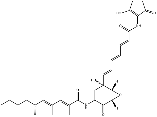
Manumycin A is an antibiotic that acts as a potent and selective farnesyltransferase (FTase) inhibitor with anti-
tumor activity. It inhibits rat brain FTase with a K
i value of 1.2 μM, thereby preventing Ras activation which requires farnesylation at the C-
terminus for membrane attachment. It exhibits significant antitumor activity against K
i-
ras-
activated solid tumors in mice at a dose of 6.3 mg/kg. Manymycin A inhibits IκB kinase (IKK), independent of FTase inhibition, in an number of cells types with effective concentrations of 2-
10 μM. In ApoE-
deficient mice, Manumycin A treatment for 22 weeks at 5 mg/kg reduced aortic fatty streak lesion size to 43% of vehicle-
treated animals, indicating FTase inhibition as a potential target for prevention or treatment of atherosclerosis.
Uses
Manumycin A is an antibiotic that acts as a potent and selective farnesyltransferase (FTase) inhibitor with anti-tumor activity.
Uses
Manumycin A from
Streptomyces parvulus has been used to inhibit IκB kinase (IKK)bnuclear κ-B essential modulator (NEMO) interaction in the homogeneous time-resolved fluorescence (HTRF)-based binding assay.
Definition
ChEBI: Manumycin A is a polyketide with formula C31H38N2O7 initially isolated from Streptomyces parvulus as a result of a random screening program for farnesyl transferase (FTase) inhibitors. It is a natural product that exhibits anticancer and antibiotic properties. It has a role as an EC 1.8.1.9 (thioredoxin reductase) inhibitor, an EC 2.5.1.58 (protein farnesyltransferase) inhibitor, an antineoplastic agent, an apoptosis inducer, an antimicrobial agent, a bacterial metabolite, an antiatherosclerotic agent and a marine metabolite. It is a polyketide, an enamide, an epoxide, an organic heterobicyclic compound, a secondary carboxamide and a tertiary alcohol.
Biochem/physiol Actions
Manumycin A is a natural monomeric epoxyquinoid. It has an ability to inhibit tumor necrosis factor (TNF) induced IκB kinase (IKK) activity in various cell types. In addition, manumycin A exhibits anti-tumor property by inhibiting farnesylation of oncogenic Ras.
in vivo
Manumycin A (5 mg/kg; once every two days; 2 weeks; i.p.) has anti-tumor effect in triple negative breast cancer mice[3].
Manumycin A (3 μg; once; i.m.) can correct abnormal splicing of Clcn1 in type 1 myotonic dystrophy (DM1) mice[5].
| Animal Model: | Female nude mice with breast cancer transplanted with MDAMB-231 cells[3]. |
| Dosage: | 5 mg/kg |
| Administration: | Intraperitoneal injection (i.p.); Once every two days; 2 weeks |
| Result: | Inhibited tumor growth and increased levels of ER stress markers such as Bip, CHOP, LC3, and p62 proteins, as well as higher molecular weight ubiquitinated proteins. |
| Animal Model: | HSALR transgenic mice[5]. |
| Dosage: | 3 μg |
| Administration: | Intramuscular injection (i.m.); once |
| Result: | Reduced the content of Clcn1 exon 7A introns. |
References
[1] M HARA. Identification of Ras farnesyltransferase inhibitors by microbial screening.[J]. Proceedings of the National Academy of Sciences of the United States of America, 1993, 90 6: 2281-2285. DOI:
10.1073/pnas.90.6.2281[2] A DI PAOLO. Manumycin inhibits ras signal transduction pathway and induces apoptosis in COLO320-DM human colon tumourcells[J]. British Journal of Cancer, 2000, 82 4: 905-912. DOI:
10.1054/bjoc.1999.1018[3] CHRISTOPH ARENZ DIPL.-CHEM. Manumycin A and Its Analogues Are Irreversible Inhibitors of Neutral Sphingomyelinase[J]. ChemBioChem, 2001, 2 2: 141-143. DOI:
10.1002/1439-7633(20010202)2:2<141::aid-cbic141>3.0.co;2-p[4] VIVEK SHARMA. Farnesyltransferase inhibitor manumycin targets IL1β-Ras-HIF-1α axis in tumor cells of diverse origin.[J]. Inflammation, 2012, 35 2: 516-519. DOI:
10.1007/s10753-011-9340-6[5] BANISHREE SAHA Dipankar N. Farnesyltransferase inhibitors reduce ras activation and ameliorate acetaminophen-induced liver injury in mice†[J]. Hepatology, 2009, 50 5: 1547-1557. DOI:
10.1002/hep.23180[6] P K SINGHA. Manumycin A inhibits triple-negative breast cancer growth through LC3-mediated cytoplasmic vacuolation death[J]. Cell Death & Disease, 2013, 4 1: e457-e457. DOI:
10.1038/cddis.2012.192