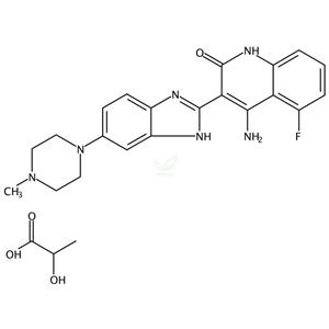
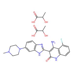
| 名称 | Dovitinib Dilactic Acid |
| 描述 | Dovitinib Dilactic Acid (Dovitinib (TKI-258) Dilactic Acid) is the Dilactic acid of Dovitinib, which is a multitargeted RTK inhibitor, mostly for class III (FLT3/c-Kit) with IC50 of 1 nM/2 nM, also potent to class IV (FGFR1/3) and class V (VEGFR1-4) RTKs with IC50 of 8-13 nM, less potent to InsR, EGFR, c-Met, EphA2, Tie2, IGFR1 and HER2. Phase 4. |
| 细胞实验 | Cell viability is assessed by 3-(4,5-dimethylthiazol)-2,5-diphenyl tetrazolium (MTT) dye absorbance. Cells are seeded in 96-well plates at a density of 5 × 10³ (B9 cells) or 2 × 10⁴ (MM cell lines) cells per well. Cells are incubated with 30 ng/mL aFGF and 100 μg/mL heparin or 1% IL-6 where indicated and increasing concentrations of Dovitinib. For each concentration of Dovitinib, 10 μL aliquots of drug or DMSO diluted in culture medium is added. For drug combination studies, cells are incubated with 0.5 μM dexamethasone, 100 nM Dovitinib, or both simultaneously where indicated. To evaluate the effect of Dovitinib on growth of MM cells adherent to BMSCs, 104 KMS11 cells are cultured on BMSC-coated 96-well plates in the presence or absence of Dovitinib. Plates are incubated for 48 to 96 hours. For assessment of macrophage colony-stimulating factor (M-CSF)-mediated growth, 5 × 10³ M-NFS-60 cells/well are incubated with serial dilutions of Dovitinib with 10 ng/mL M-CSF and without granulocyte-macrophage colony-stimulating factor (GM-CSF). After 72 hours cell viability is determined using Cell Titer-Glo Assay. Each experimental condition is performed in triplicate. (Only for Reference) |
| 激酶实验 | In vitro kinase assays: The inhibitory concentration of 50% (IC50) values for the inhibition of RTKs by Dovitinib are determined in a time-resolved fluorescence (TRF) or radioactive format, measuring the inhibition by Dovitinib of phosphate transfer to a substrate by the respective enzyme. The kinase domains of FGFR3, FGFR1, PDGFRβ, and VEGFR1-3 are assayed in 50 mM HEPES (N-2-hydroxyethylpiperazine-N′-2-ethanesulfonic acid), pH 7.0, 2 mM MgCl2, 10 mM MnCl2 1 mM NaF, 1 mM dithiothreitol (DTT), 1 mg/mL bovine serum albumin (BSA), 0.25 μM biotinylated peptide substrate (GGGGQDGKDYIVLPI), and 1 to 30 μM adenosine triphosphate (ATP) depending on the Km for the respective enzyme. ATP concentrations are at or just below Km. For c-KIT and FLT3 reactions the pH is raised to 7.5 with 0.2 to 8 μM ATP in the presence of 0.25 to 1 μM biotinylated peptide substrate (GGLFDDPSYVNVQNL). Reactions are incubated at room temperature for 1 to 4 hours and the phosphorylated peptide captured on streptavidin-coated microtiter plates containing stop reaction buffer (25 mM EDTA [ethylenediaminetetraacetic acid], 50 mM HEPES, pH 7.5). Phosphorylated peptide is measured with the DELFIA TRF system using a Europium-labeled antiphosphotyrosine antibody PT66. The concentration of Dovitinib for IC50 is calculated using nonlinear regression with XL-Fit data analysis software version 4.1 (IDBS). Inhibition of colony-stimulating factor-1 receptor (CSF-1R), PDGFRα, insulin receptor (InsR), and insulin-like growth factor receptor 1 (IGFR1) kinase activity is determined at ATP concentrations close the Km for ATP. |
| 体外活性 | Dovitinib 对 FGF 刺激下生长的 WT 和 F384L-FGFR3 表达的 B9 细胞表现出强力抑制作用,具有 25 nM 的 IC50 值。此外,Dovitinib 还能抑制表达各种活化突变型 FGFR3 的 B9 细胞的增殖。有趣的是,对 Dovitinib 的敏感性在不同 FGFR3 突变之间的差异很小,IC50 值介于 70 到 90 nM 之间。仅含有载体的 IL-6 依赖性 B9 细胞(B9-MINV)对 Dovitinib 的抑制活性在高达 1 μM 的浓度下表现出抵抗性。Dovitinib 抑制 KMS11(FGFR3-Y373C)、OPM2(FGFR3-K650E)和 KMS18(FGFR3-G384D)细胞的细胞增殖,IC50 值分别为 90 nM(KMS11 和 OPM2)和 550 nM。Dovitinib 抑制 FGF 介导的 ERK1/2 磷酸化并在表达 FGFR3 的原发性 MM 细胞中引起细胞毒性。与未共培养 BMSCs 的细胞相比,BMSCs 能为用 500 nM Dovitinib 处理的细胞提供轻微的抗性,前者的生长抑制率为 44.6%,而后者为 71.6%。Dovitinib 抑制由 M-CSF 驱动生长的小鼠骨髓增生性细胞系 M-NFS-60 的增殖,其 EC50 为 220 nM。[1] SK-HEP1 细胞对 Dovitinib 的处理导致细胞数量剂量依赖性下降,G2/M 期阻滞,G0/G1 和 S 期减少,抑制无锚生长,并阻断 bFGF 引起的细胞运动。在 SK-HEP1 细胞中,Dovitinib 的 IC50 值约为 1.7 μM。Dovitinib 同时显著减少了 SK-HEP1 和 21-0208 细胞中 FGFR-1、FGFR底物 2α(FRS2-α)和 ERK1/2 的基础磷酸化水平,但对 Akt 无影响。在 21-0208 HCC 细胞中,Dovitinib 显著抑制了 bFGF 引起的 FGFR-1、FRS2-α、ERK1/2 的磷酸化,但对 Akt 无影响。[2] |
| 体内活性 | Dovitinib在体内诱导细胞静止和细胞毒性反应,导致表达FGFR3的肿瘤减小。[1] Dovitinib通过剂量和暴露程度依赖的方式,抑制肿瘤异种移植物中表达的目标受体酪氨酸激酶(RTKs)。Dovitinib强效地抑制六种HCC细胞系的肿瘤生长。抑制血管生成与FGFR/PDGFRβ/VEGFR2信号通路的失活相关。在原位模型中,Dovitinib强效地抑制原发性肿瘤生长和肺转移,并显著延长小鼠的存活时间。[2] Dovitinib的使用显著抑制肿瘤生长并导致肿瘤回退,包括大型、已建立的肿瘤(500-1,000 mm3)。[3] |
| 存储条件 | Powder: -20°C for 3 years | In solvent: -80°C for 1 year | Shipping with blue ice/Shipping at ambient temperature. |
| 溶解度 | DMSO : 83 mg/mL (144.96 mM), Sonication is recommended.
H2O : 64 mg/mL (111.77 mM), Sonication is recommended.
Ethanol : <1 mg/mL
|
| 关键字 | VEGFR3/FLT4 | FLT3 | FGFR3 | FGFR1 | Dovitinib Dilactic Acid | c-Kit | cKit |
| 相关产品 | glycine | Amlexanox | Gilteritinib | Lenvatinib | Formononetin | Ferulic Acid | Regorafenib | Pazopanib | Dasatinib | Ethyl cinnamate | Sorafenib | Nintedanib esylate |


