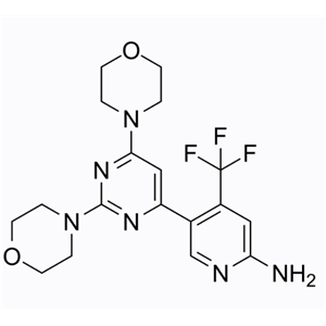
名称 | Buparlisib |
描述 | Buparlisib (BKM120) is an orally bioavailable specific oral inhibitor of the pan-class I PI3K (IC50s: 52/166/116/nM for p110α, p110β, and p110δ). |
细胞实验 | A2780 cells were cultured in DMEM supplemented with 10% FBS. L-glutamine, sodium pyruvate, and antibiotics. Cells were plated in the same medium at a density of 1000 cells per well, 100 ul per well into black-walled-clear-bottom plates and incubated for 3-5 hours. Test compounds supplied in DMSO (20 mM) were diluted further into DMSO (7.5 ul of 20 mM test compound in 22.5 ul DMSO. Mix well, transfer 10 ul to 20 ul DMSO, repeat until 9 concentrations have been made). The diluted test compound solution (2uL), was then added to the cell medium (500 ul) cell medium. Equal volumes of this solution (100 uL) were added to the cells in 96 well plates and incubated at 37 oC for 3 days and developed using Cell Titer Glo. Inhibition of cell proliferation was determined by luminescence read using Trilux [1]. |
激酶实验 | Compounds to be tested were dissolved in DMSO and directly distributed into a black 384-well plate at 1.25 μL per well. To start the reaction, 25 μL of 10 nM PI3 kinase and 5 μg/mL 1-alpha-phosphatidylinositol (PI) in assay buffer (10 mM Tris pH 7.5, 5 mM MgCl2, 20 mM NaCl, 1 mM DTT and 0.05% CHAPS) were added into each well followed by 25 μL of 2 μM ATP in assay buffer. The reaction was performed until approx 50% of the ATP was depleted, and then stopped by the addition of 25 μL of KinaseGlo solution. The stopped reaction was incubated for 5 minutes and the remaining ATP was then detected via luminescence [1]. |
动物实验 | Six- to eight-week-old female severe combined immunodeficiency (SCID) mice were housed and monitored in the MD Anderson Cancer Center animal research facility. SCID mice were subcutaneously inoculated in the right flank with 1 million ARP-1 or MM.1S cells suspended in 50 μl phosphate-buffered saline (PBS). After palpable tumor developed (tumor diameter ≥5 mm), mice were treated with intraperitoneal injection of DMSO/PBS or BKM120 (5 μM per kg per day) for 15 days. Tumor sizes were measured every 5 days, and blood samples were collected at the same period. Tumor burdens were evaluated by measuring tumor size and detecting the circulating human kappa chain or lambda chain [3]. |
体外活性 | Buparlisib对类I PI3K(包括常见的p110R突变体)表现出50-300 nM的活性。此外,其对类III和类IV PI3K的抑制活性较低,分别观察到VPS34、mTOR、DNAPK和PI4K的生化活性在2、5、>5和>25 μM。在所有细胞系中,通路调节和抗增殖活性与细胞内PI3K抑制一致[1]。Buparlisib在11种人类胃癌细胞系中展现出抗增殖活性,通过减少mTOR下游信号传导。但是Buparlisib处理通过稳定胰岛素受体底物-1来取消反馈抑制,从而增加了p-AKT。在KRAS突变型胃癌细胞中,处理Buparlisib后,p-ERK或p-STAT3也有所增加[2]。BKM120在多个骨髓瘤(MM)细胞系和新鲜分离的初级MM细胞中诱导细胞生长抑制和凋亡。此外,BKM120与地塞米松在对地塞米松敏感的MM细胞中显示出协同细胞毒性。低剂量的BKM120和地塞米松,各自独立具有有限的细胞毒性,能在MM.1S和ARP-1中诱导显著的细胞凋亡。BKM120暴露导致通过上调p27 (Kip1)和下调cyclin D1来引发细胞周期阻滞,并通过下调抗凋亡的XIAP和上调表达细胞毒性小型异构体BimS来诱发依赖于半胱天冬酶的凋亡[3]。 |
体内活性 | 在A2780异种移植瘤模型中,口服Buparlisib以3、10、30、60及100 mg/kg剂量对pAKTSer473进行了剂量依赖性调节。在3及10 mg/kg剂量时观察到pAKTser473的部分抑制;而在30、60或100 mg/kg剂量时,则观察到几乎完全抑制。pAKT的抑制与血浆及肿瘤内化合物暴露程度密切相关。pAKT调节同样依赖于时间,当血浆和肿瘤暴露量大约为2 μM时,在10小时时间点,60及100 mg/kg剂量实现了>90%的靶点调节[1]。与对照组小鼠相比,每天以5 μM/kg剂量Buparlisib治疗的小鼠显示出明显较小的肿瘤负担,此负担通过肿瘤体积和循环人类κ链水平来衡量。此外,Buparlisib治疗显著延长了负载瘤小鼠的生存期[3]。 |
存储条件 | Powder: -20°C for 3 years | In solvent: -80°C for 1 year | Shipping with blue ice. |
溶解度 | DMSO : 76 mg/mL (185.2 mM) H2O : Insoluble Ethanol : 2 mg/mL
|
关键字 | Phosphoinositide 3-kinase | inhibit | BKM 120 | Buparlisib | Inhibitor | PI3K | NVP-BKM-120 | BKM-120 | Apoptosis | NVP-BKM 120 |
相关产品 | L-Glutamic acid | Metronidazole | 5-Fluorouracil | Dextran sulfate sodium salt (MW 4500-5500) | Stavudine | Tributyrin | Myricetin | Sorafenib | L-Ascorbic acid | Acetylcysteine | Salicylic acid | Sodium 4-phenylbutyrate |
相关库 | 抑制剂库 | 经典已知活性库 | 抗癌活性化合物库 | 已知活性化合物库 | 自噬库 | 激酶抑制剂库 | 抗衰老化合物库 | 药物功能重定位化合物库 | 抗癌临床化合物库 | 抗癌药物库 |