
7YR企业会员
发布人:爱必信(上海)生物科技有限公司
发布日期:2026/7/28 14:44:40

心肌梗死(MI)是全球高发病率、高死亡率的心血管疾病。心梗后巨噬细胞介导的过度炎症反应是造成心肌细胞损伤、心脏修复受阻、心室不良重构的核心诱因。目前临床上仍缺乏针对心梗炎症的有效靶向药物,因此挖掘巨噬细胞炎症调控的关键分子、解析信号通路,是开发心梗治疗方案的核心方向。
已有研究证实RNA结合蛋白广泛参与免疫调控,ILF3(白细胞介素增强子结合因子3)在天然免疫、肿瘤、慢性血管疾病中发挥作用,但它在心梗急性炎症中的功能与分子机制尚不明确。基于此,本团队围绕巨噬细胞ILF3展开系统性探索。
整篇文章遵循表型验证→功能验证→机制深挖→靶点验证的经典科研逻辑,实验设计严谨、环环相扣:
表达筛选:结合转录组数据库,筛选心梗及巨噬细胞促炎极化过程中差异表达的RNA结合蛋白,锁定ILF3;在急性心梗(AMI)患者、心梗小鼠模型中验证ILF3的表达分布与表达时序。
功能验证:构建巨噬细胞特异性ILF3敲除小鼠(ILF3-cKO)与过表达小鼠(ILF3-cTG),从心脏功能、心肌凋亡、炎症水平、组织修复四大维度,明确ILF3促心梗损伤的生物学功能。
机制探究:利用蛋白质组、IP-MS筛选ILF3互作蛋白,锁定下游靶蛋白HNRNPA2B1;解析ILF3调控HNRNPA2B1的泛素化修饰机制,再延伸至m6A甲基化、mRNA稳定性、下游信号通路。
转化验证:靶向抑制通路关键分子(HNRNPA2B1、Irak4),在动物模型中验证治疗效果,评估临床转化潜力。
研究发现,无论是急性心梗患者还是小鼠心梗模型,心脏梗死区、外周血单核细胞(PBMC)中ILF3表达均显著上调。
小鼠心梗模型中,ILF3表达在术后第3天达到峰值(对应心梗早期炎症高峰期),4周后回落至基线水平(原文图1C);
免疫荧光证实,ILF3主要富集于招募型CCR2⁺巨噬细胞,心肌细胞、成纤维细胞、中性粒细胞中几乎无明显上调(原文图1E、1H、1I);
体外实验中,LPS/IFNγ诱导巨噬细胞促炎极化后,ILF3表达24h达到峰值(原文图1L)。
结论:心梗早期炎症阶段,巨噬细胞是ILF3的主要表达细胞,提示ILF3与巨噬细胞促炎表型高度相关。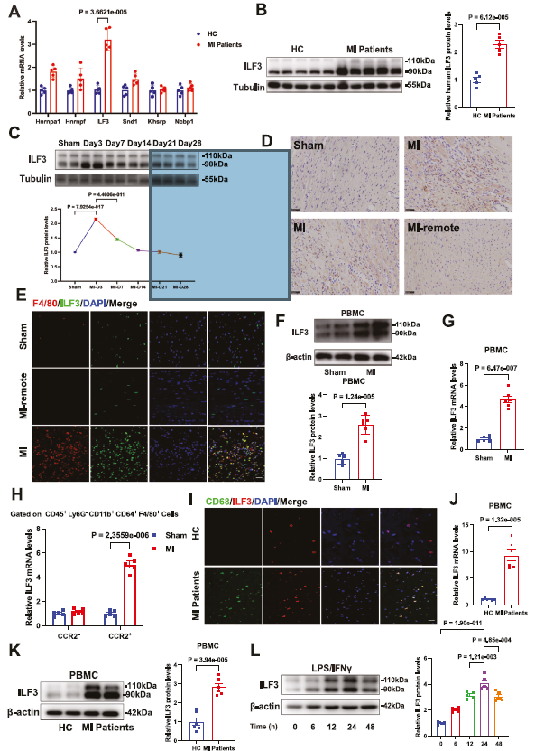 Fig. 1 Macrophages highly expressing ILF3 infiltrate murine and human AMI tissues. A Analysis of Hnrnpa1, Hnrnpf, ILF3, Snd1, Khsrp and Ncbp1 mRNA expression in cardiac tissue from patients with AMI and healthy controls (n = 5). B Immunoblot analysis of ILF3 levels in the infarct zone of patients with AMI and the corresponding cardiac tissue of healthy controls (n = 5). C Immunoblot analysis of ILF3 levels in murine infarct hearts at multiple days post-MI (n = 6). D Immunohistochemical staining of ILF3 in heart tissues after sham surgery or 3 days post-MI (scale bar = 50 μm; n = 5). E Immunofluorescence costaining of F4/80 (red) or ILF3 (green) in cardiac tissues after sham surgery or 3 days post-MI (scale bar = 20 μm; n = 6). F Immunoblot analysis of ILF3 levels in PBMCs from sham or MI mice (n = 6). G Analysis of ILF3 mRNA expression in PBMCs from sham or MI mice (n = 6). H ILF3 mRNA levels in CCR2- and CCR2+ cardiac macrophage subsets from WT hearts before and 3 days after MI (n = 5). I Immunofluorescence costaining of CD68 (green) and ILF3 (red) in heart tissues from patients with AMI and healthy controls (scale bar = 20 μm; n = 5). J ILF3 mRNA expression levels in PBMCs from healthy controls and patients with AMI (n = 6). K Western blotting and quantification of ILF3 in PBMCs from healthy controls and patients with AMI (n = 6). L Western blotting and quantification of ILF3 expression in BMDMs stimulated with LPS/IFNγ (200 ng/ml or 40 ng/ml) for 0, 6, 12, 24, or 48 h (n = 5). Data were analyzed using an unpaired two-tailed Student’s t test (B, F, G, J, K), multiple unpaired t tests with Benjamini and Hochberg false discovery rate FDR) correction (A), one-way ANOVA with Bonferroni multiple comparison tests (C, L) and two-way ANOVA with Bonferroni multiple comparison tests (H)
利用巨噬细胞特异性敲除小鼠开展体内功能实验,获得多项关键阳性结果:
心脏功能提升:心梗21天后,ILF3-cKO小鼠左室射血分数(EF)、短轴缩短率(FS)显著升高,心室腔扩张得到抑制,心脏肥厚减轻(原文图2A、2B);
梗死面积与纤维化减少:TTC染色显示梗死面积缩小,Masson、天狼星红染色证实心肌纤维化面积降低,瘢痕结构更稳定(原文图2C、2D);
生存率提升:ILF3-cKO小鼠心梗后21天生存率(83%)远高于野生型小鼠(67%),有效降低心脏破裂风险(原文图2F);
心肌凋亡减少:TUNEL染色显示梗死边界区心肌细胞凋亡数量大幅下降(原文图3A)。
结论:巨噬细胞ILF3是心梗后心肌损伤、心室重构的有害分子,敲除ILF3可显著保护心脏。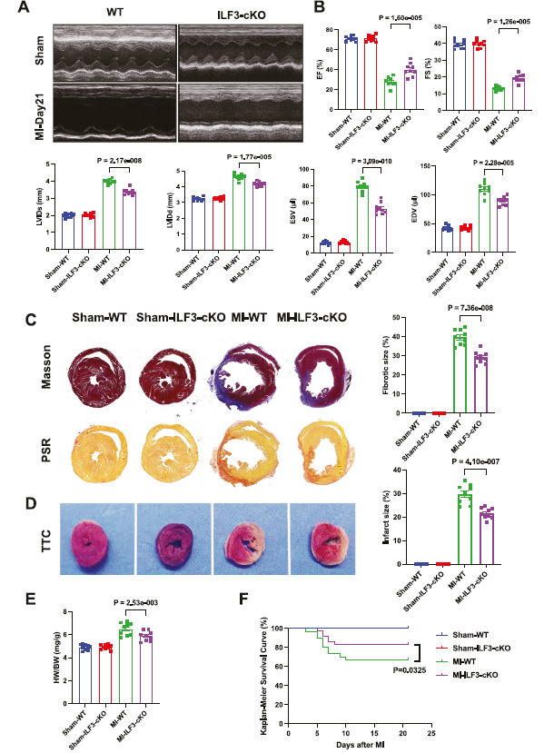 Fig. 2 ILF3 deficiency in macrophages ameliorates myocardial dysfunction and adverse remodeling and improves post-MI survival.
A Representative M-mode echocardiograms obtained from WT and ILF3-cKO mice on day 0 and day 21 after MI (n = 9). B EF, FS, LVIDs, LVIDd,ESV, and EDV in WT and ILF3-cKO mice. C Masson’s Trichrome (MT) and Picrosirius Red (PSR) staining of transverse cross-sections of hearts obtained from WT and ILF3-cKO mice on day 21 after MI, and the size of fibrosis was quantified (n = 9). D TTC staining of transverse crosssections of hearts obtained from WT and ILF3-cKO mice on day 3 after MI, and the infarct size was quantified (n= 9). E Ratio of heart weight to body weight (HW/BW) of mice on day 21 after MI (n = 9). F Survival analysis of WT (n = 30) and ILF3-cKO mice (n = 30) after MI or sham operation (n = 20). The data were analyzed using two-way ANOVA with the Bonferroni correction for multiple comparisons (B, C, D, E). Survival rates were analyzed using the Kaplan‒Meier method and compared using the log-rank test (F)
抑制促炎表型:ILF3敲除后,梗死区促炎基因(iNOS、TNF-α、IL-6)表达下调,M1型促炎巨噬细胞(iNOS⁺)数量减少(原文图3B、3C);
促进抗炎修复表型:抗炎基因(CD206、IL-10、Arg1)表达升高,M2型修复型巨噬细胞(Arg1⁺)富集(原文图3D、3F);
助力组织修复:心梗修复期,ILF3缺失促进肌成纤维细胞增殖、胶原沉积与血管新生(CD31⁺内皮细胞增多),加速心肌修复(原文图3I、3J、3K)。
结论:ILF3推动巨噬细胞向促炎表型极化,阻断炎症消退、抑制心脏修复;敲除ILF3可平衡炎症稳态。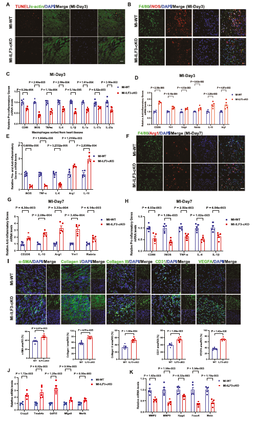 Fig. 3 ILF3 deficiency in macrophages mitigates apoptosis, limits the inflammatory response and enhances post-MI cardiac repair. A TUNEL (red)-α-actin (green) staining of border infarct heart tissues from WT and ILF3-cKO mice (scale bar = 20 μm; n = 5). B Immunofluorescence costaining of F4/80 (green) and iNOS (red) in infarcted heart tissues from WT and ILF3-cKO mice (scale bar = 20 μm; n = 5). C, Analysis of proinflammatory gene mRNA expression in infarcted heart tissues from WT and ILF3-cKO mice (n = 5). D Analysis of anti-inflammatory gene mRNA expression in infarcted heart tissue from WT and ILF3-cKO mice (n = 5). E Analysis of the mRNA expression of major proinflammatory and anti-inflammatory genes in macrophages from infarcted heart tissue from WT and ILF3-cKO mice (n = 5). F Immunofluorescence costaining of F4/80 (green) and Arg1 (red) in infarcted heart tissues from WT and ILF3-cKO mice (scale bar = 20 μm; n = 5). G Analysis of antiinflammatory gene mRNA expression in infarcted heart tissue from WT and ILF3-cKO mice (n = 5). H Analysis of proinflammatory gene mRNA expression in infarcted heart tissue from WT and ILF3-cKO mice (n= 5). I Immunofluorescence staining and quantification of α-SMA, collagen I, collagen III, CD31 and VEGFA expression in infarcted heart tissues from WT and ILF3-cKO mice (scale bar = 20 μm; n = 5). J Τhe mRNA expression levels of prorepair genes in infarct heart tissues from WT and ILF3-cKO mice (n = 5). K Τhe mRNA expression levels of anti-repair genes in infarct heart tissues from WT and ILF3-cKO mice (n = 5). Data were analyzed using unpaired two-tailed Student’s t tests (I) and multiple unpaired t tests with Benjamini and Hochberg false discovery rate (FDR) correction (C, D, E, G, H, J, K)
这是本文最核心的机制创新,层层解析蛋白互作、翻译后修饰、表观修饰三大调控环节:
1. ILF3结合并稳定HNRNPA2B1
蛋白质组与IP-MS筛选发现ILF3与HNRNPA2B1直接互作(原文图4C、5A-5D)。ILF3通过自身RRM1、RRM2结构域结合HNRNPA2B1,不影响其mRNA水平,仅抑制蛋白降解。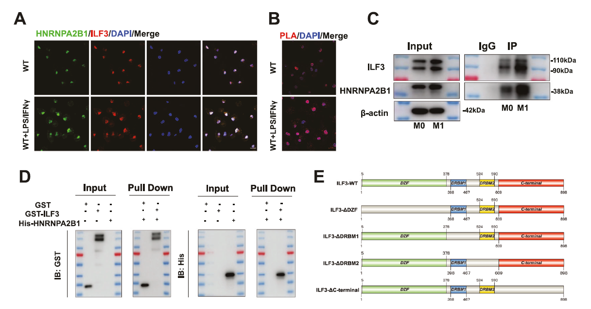 Fig. 5 ILF3 deficiency downregulates HNRNPA2B1 expression in association with ubiquitination. A Immunofluorescence costaining of HNRNPA2B1 (green) and ILF3 (red) in BMDMs with or without LPS/IFNγ treatment (scale bar = 10 μm; n = 5). B PLA staining (red) of HNRNPA2B1 and ILF3 in BMDMs with or without LPS/IFNγ treatment (scale bar = 10 μm; n = 5). C Co-IP assay and visualization of interactions between ILF3 and HNRNPA2B1 in BMDMs from WT mice with or without LPS/IFNγ treatment (n = 4). D Results of a GST pull-down assay and visualization of interactions between the His-HNRNPA2B1 protein and the purified GST-ILF3 protein (n = 4). E Schematic diagram of ILF3 and its deletion mutants.
2. ILF3阻断Trim21介导的K48泛素化
E3泛素连接酶Trim21可靶向HNRNPA2B1第112位赖氨酸(K112),介导K48连接的泛素化降解;而ILF3与HNRNPA2B1的结合会空间位阻K112位点,阻止Trim21识别并降解HNRNPA2B1(原文图5J、6A-6K)。ILF3敲除后,HNRNPA2B1泛素化水平升高、蛋白量下降。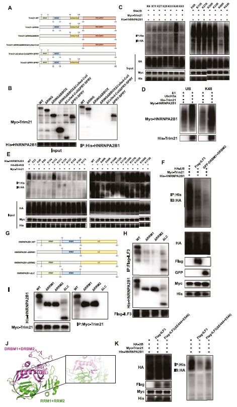 Fig. 6 ILF3 inhibits K48-linked polyubiquitination by trim21 targeting K112 of HNRNPA2B1. A Schematic diagram of Trim21 and its deletion mutants. B Co-IP assay in HEK293T cells between His-HNRNPA2B1 and deletion mutants of Flag-ILF3. C Co-IP assay of HNRNPA2B1 ubiquitination in HEK293T cells transfected with Myc-Trim21, His-HNRNPA2B1, HA-UB and mutant ubiquitin. D In vitro HNRNPA2B1 ubiquitination assay with purified recombinant proteins, including Myc-HNRNPA2B1 and His-Trim21, in the presence of E1, E2 (UbcH5a), UB, and UB (K48). E Co-IP assay of the polyubiquitination of His-HNRNPA2B1 and its mutants in HEK293T cells cotransfected with HA-K48 and Myc-Trim21. F Co-IP assay of HNRNPA2B1 ubiquitination in HEK293T cells transfected with Myc-Trim21, His-HNRNPA2B1, HA-UB, Flag-ILF3, and GFPDRBM1 + DRBM2. G Schematic diagram of HNRNPA2B1 and its deletion mutants. H Co-IP assay in HEK293T cells between Flag-ILF3 and His-HNRNPA2B1 deletion mutants. I Co-IP of Myc-Trim21 and His-HNRNPA2B1 deletion mutants in HEK293T cells. J Molecular docking results of DRBM1 + DRBM2 of ILF3 and RRM1 + RRM2 of HNRNPA2B1. K Co-IP assay of HNRNPA2B1 ubiquitination in HEK293T cells transfected with Myc-Trim21, His-HNRNPA2B1, HA-UB, Flag-ILF3 and Flag-ILF3 (ΔE444 + E546)
3. HNRNPA2B1通过m6A修饰稳定Irak4 mRNA
HNRNPA2B1是经典m6A阅读蛋白,可结合Irak4 mRNA的m6A位点,提升mRNA稳定性、上调Irak4表达(原文图7Q-7W)。ILF3缺失会下调HNRNPA2B1,进而降低Irak4水平。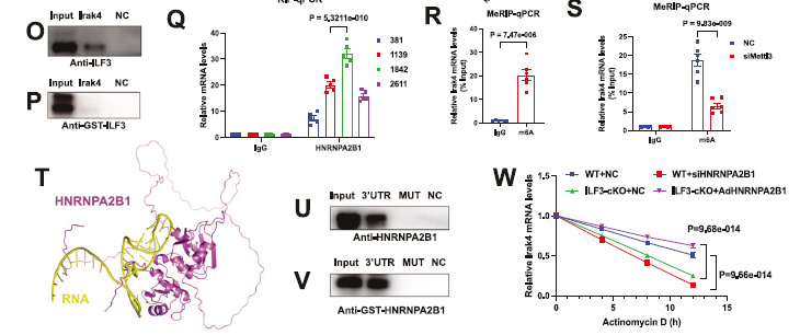 Fig. 7 The ability of the ILF3/HNRNPA2B1 axis to upregulate Irak4 expression depends on m6A modification. O Western blotting of ILF3 after the RNA pull-down assay in BMDMs; the NC served as a negative control (n=3). PWestern blotting of GST-ILF3 after the RNA pull-down assay (n=3). Q RIP assay for enrichment of Irak4mRNA at 381, 1139,1842, and 2611 sites with HNRNPA2B1 in BMDMs (n=5). R MeRIP assay for m6A enrichment of Irak4mRNA in BMDMs (n=6). S MeRIP assay for m6A enrichment of Irak4mRNA in BMDMs transfected with NC or siMettl3 (n=6). T Molecular docking results of HNRNPA2B1 and Irak4 mRNA. U Western blotting of HNRNPA2B1 after an RNA pull-down assay with the Irak4 3’UTR with or without m6A site mutation in BMDMs. V Western blotting of GSTHNRNPA2B1
after an RNA pull-down assay with the Irak4 3’UTR with or without m6A sitemutation.WRelative Irak4 remaining RNA levels at different
time points after actinomycin D treatment in BMDMs transfected with siHNRNPA2B1 from WT mice and with AdHNRNPA2B1 from ILF3-cKO mice (n=4). Data were analyzed using an unpaired two-tailed Student’s t test (C, D, L, M, R), multiple unpaired t tests with Benjamini and Hochberg false discovery rate (FDR) correction (K) and two-way ANOVA with Bonferroni multiple comparison tests (E, F, H, I, J, N, Q, S, W)
4. 下游通路激活与炎症放大
Irak4进一步激活c-jun/c-fos炎症通路,放大心梗早期炎症反应,最终加重心肌损伤与不良重构(原文图7A、7B)。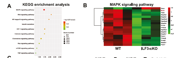
Fig. 7 The ability of the ILF3/HNRNPA2B1 axis to upregulate Irak4 expression depends on m6A modification. A Bubble diagram showing the different signaling pathways identified by KEGG enrichment analysis. B Heatmap showing the upregulated and downregulated proteins involved in theMAPK signaling pathway.
完整通路总结:心梗后巨噬细胞ILF3上调→结合HNRNPA2B1并抑制其Trim21介导的K48泛素化降解→HNRNPA2B1累积→通过m6A修饰稳定Irak4 mRNA→Irak4高表达激活c-jun/c-fos通路→巨噬细胞过度促炎→心肌损伤、修复受阻。
在巨噬细胞ILF3过表达(ILF3-cTG)心梗小鼠中,分别采用敲低HNRNPA2B1、口服Irak4抑制剂Zimlovisertib两种干预手段:
两种方案均能逆转ILF3过表达导致的心脏功能恶化,减少心肌纤维化(原文图8A-8C);
急性期抑制炎症、减少心肌凋亡,修复期促进血管新生与胶原沉积(原文图8D-8F)。
转化价值:HNRNPA2B1、Irak4可作为心梗炎症的潜在药物靶点,Irak4抑制剂Zimlovisertib展现出心血管疾病应用前景。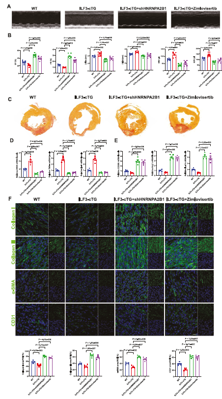 Fig. 8 Targeting HNRNPA2B1 and Irak4 effectively improved cardiac function and myocardial injury and promoted cardiac repair post-
MI in ILF3-cTG mice. A Representative M-mode echocardiograms obtained from WT and ILF3-cTG mice on day 21 after MI (n = 7). B EF, FS, LVIDs, LVIDd, ESV, and EDV in WT and ILF3-cTG mice (n = 7). C PSR staining of transverse cross-sections of hearts obtained from WT and ILF3-cTG mice on day 21 after MI (n = 7). D Analysis of proinflammatory gene mRNA expression in infarcted heart tissues from WT and ILF3-cTG mice on day 3 after MI (n = 5). E Analysis of anti-inflammatory gene mRNA expression in infarcted heart tissue from WT and ILF3-cTG mice on day 7 after MI (n = 5). F Immunofluorescence staining and quantification of Collagen I, Collagen III, α-SMA, and CD31 expression in infarcted heart tissue from WT and ILF3-cTG mice on day 7 after MI (scale bar = 20 μm; n = 5). The data were analyzed using two-way ANOVA with the Bonferroni multiple comparison test (B, D, E, F)
本研究依托两款Absin核心产品完成关键样本前处理实验,保障流式细胞分析、细胞分选等实验数据准确可靠,具体应用如下:
应用场景
用于心梗小鼠心脏组织解离,制备单细胞悬液,支撑巨噬细胞亚群流式分析、原代细胞分选实验。
产品作用
优化复合酶解体系,可温和解离病变心肌组织,完整保留巨噬细胞表面抗原,避免抗原降解造成流式检测误差;
解离后细胞活率高、碎片少,无需二次纯化,直接适配流式上机与流式分选,适配心梗坏死+纤维化混合组织的解离需求。
应用场景
心脏组织酶解后,对细胞悬液进行过滤筛分,去除组织团块与杂质,制备合格单细胞悬液,为后续流式检测、细胞分选提供标准样本。
产品作用
标准70μm孔径,有效截留未充分解离的组织块、细胞聚集体,获得单一细胞悬液;
筛网材质稳定,通透性好,过滤效率高,可有效降低流式管路堵塞风险,保证细胞分群与分选结果稳定。
科学创新:本研究首次确立巨噬细胞ILF3是心梗后炎症与心肌损伤的关键驱动分子,打通“蛋白泛素化-m6A表观修饰-炎症通路”的全新调控轴,补充了心血管免疫领域的分子机制空白;
临床价值:证实HNRNPA2B1、Irak4为有效药物靶点,Irak4抑制剂Zimlovisertib在心梗模型中展现治疗效果,为缺血性心脏病新药研发提供新思路;
实验亮点:体内外模型结合、多技术联用(转录组、蛋白质组、泛素化、m6A、流式、病理染色),实验体系完整严谨。
结合本文实验场景,针对心肌梗死、动脉粥样硬化、心肌炎等心血管免疫研究,优先推荐以下Absin明星产品:
相关新闻资讯
