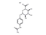
| Name | Acetaminophen |
| Description | Acetaminophen (APAP) is a COX inhibitor that inhibits COX-1 and COX-2 (IC50=113.7/25.8 μM). Acetaminophen has antipyretic and analgesic activity as well as weak anti-inflammatory activity. |
| Cell Research | Cells are exposed to Acetaminophen for 48 hours. Cell viability is determined by the trypan blue exclusion method. Intracellular GSH is measured by recording the disulfide, GS-TNB and 5-thio-nitrobenzoic acid (TNB), the yellow colored compound formed by the reaction between GSH with DTNB.(Only for Reference) |
| Kinase Assay | Effect of inhibition of Acetaminophen on COX-1 and COX-2 activity in human whole blood: For COX-1 assay, aliquots of human whole blood drawn from healthy volunteers without anticoagulant are transferred to glass tubes containing Acetaminophen or DMSO, serum is separated by centrifugation after clotting, and serum TxB2 levels are determined. For COX-2 assay, aliquots of heparinized whole blood are incubated with LPS (10 μg/mL) and aspirin (10 μg/mL), plus Acetaminophen or DMSO for 24 hours at 37 °C, plasma is separated by centrifugation, and PGE2 levels are determined subsequently. The degree of COX-1 or COX-2 inhibition is calculated as the percentage change of plasma eicosanoid (TxB2 for COX-1 and PGE2 for COX-2).Concentration response curves are fitted by a sigmoidal regression with variable slope for both enzymatic assays, and the 50% inhibitory concentration (IC50) values are derived by using of PRISM Version 3.0. |
| In vitro | METHODS: Non-melanoma and melanoma cell lines were treated with Acetaminophen (100 µM) for 48 h and cell viability was measured using MTT Assay.
RESULTS: Acetaminophen showed considerable toxicity in melanoma cell lines SK-MEL-5, MeWo, B16-F0 and B16-F10, resulting in 40±3%, 45±7%, 66±8% and 60±5% cytotoxicity, respectively. Acetaminophen showed negligible toxicity at 100 μM in the non-melanoma cell lines PC-3, BJ, Saos-2 and SW-620 cells. [1]
METHODS: Neuroblastoma cells SH-SY5Y were treated with Acetaminophen (2 mM) for 24-48 h, and the expression levels of target proteins were detected using Western Blot.
RESULTS: Acetaminophen induced cytochrome c release from mitochondria in a time-dependent manner, reaching a maximum level after 48 h of treatment. In addition, immunoblot analysis of cytoplasmic and mitochondrial fractions showed that Acetaminophen was able to induce the accumulation of Bax into mitochondria at 24 h after treatment. [2] |
| In vivo | METHODS: To detect hepatotoxicity in vivo, Acetaminophen was administered intraperitoneally to mice (300 mg/kg) and rats (1 g/kg).
RESULTS: Extensive liver necrosis was observed in mice, but little damage was observed in rat samples. The rats were highly resistant to Acetaminophen-induced liver injury. [3]
METHODS: To test for in vivo hepatotoxicity, Acetaminophen was administered to aged and weakened mice acutely (300 mg/kg by gavage), chronically (100 mg/kg by diet once daily for six weeks), or subacutely (250 mg/kg by gavage three times daily for three days).
RESULTS: There was no overall increase in Acetaminophen hepatotoxicity with age or frailty in mice, despite changes in certain pathways that would be expected to affect susceptibility to Acetaminophen toxicity. [4] |
| Storage | The compound is unstable in solution. Please use soon | Powder: -20°C for 3 years | Shipping with blue ice/Shipping at ambient temperature. |
| Solubility Information | DMSO : 237 mg/mL (1567.88 mM), Sonication is recommended.
H2O : 20 mg/mL (132.31 mM), Sonication is recommended. The compound is unstable in solution, please use soon.
Ethanol : 15.1 mg/mL (99.89 mM), Sonication is recommended.
|
| Keywords | Inhibitor | inhibit | HistoneAcetyltransferase | Histone Acetyltransferase | HATs | HAT | EndogenousMetabolite | Endogenous Metabolite | Cyclooxygenase | COX-2 | COX-1 | COX | Acetaminophen |
| Inhibitors Related | Sucrose | Aceglutamide | L-Pyroglutamic acid | Emtricitabine | Levulinic acid | D(+)-Raffinose pentahydrate | D-(+)-Trehalose dihydrate | Manganese chloride (tetrahydrate) | Glycerol | Thymidine | Trometamol | Gluconate Calcium |

 United States


