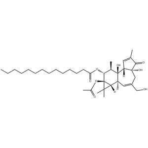
| Description | Phorbol 12-myristate 13-acetate (PMA), a phorbol ester, is a commonly used PKC activator. |
|---|
| Related Catalog | Signaling Pathways >> Epigenetics >> PKC Signaling Pathways >> TGF-beta/Smad >> PKC Research Areas >> Inflammation/Immunology Natural Products >> Others |
|---|
| Target | PKC:11.7 nM (EC50) |
|---|
| In Vitro | In order to examine the role of PKC in p38MAPK phosphorylation, the cells are stimulated with the PKC activator, PMA (100 nM), which mimics the binding of DAG, the natural activator of PKC, to the C1 region of the PKCs. p38MAPK phosphorylation by PMA is observed in the two cell types similar to that observed by GnRH in αT3-1 cells, that is, a slow sustained activation (3.2-fold and 3.6-fold, respectively at 30 min). The paradoxical findings that PKCs activated by GnRH and PMA play a differential role in p38MAPK phosphorylation may be explained by differential localization of the PKCs. Basal, GnRH- and PMA- stimulation of p38MAPK phosphorylation in αT3-1 cells is mediated by Ca2+ influx via voltage-gated Ca2+ channels and Ca2+ mobilization, while in the differentiated LβT2 gonadotrope cells it is mediated only by Ca2+ mobilization[2]. |
|---|
| In Vivo | PMA is a PKC agonist, which reverses the damage induced by 5-hydroxydecanoic acid (5-HD). Thus, activation of the mitoKATP protected mitochondrial function in SOD and MDA via the PKC pathway[3]. |
|---|
| Cell Assay | αT3-1 and LβT-2 cells are grown in monolayer cultured in DMEM supplemented with 10% fetal calf serum (FCS) and L-glutamine 2 mM, penicillin and streptomycin (100 units/mL) in humidified incubator 5% CO2 at 37°C. Serum starvation is with 0.1% FCS in the same medium for 16 h. GnRH and PMA are then added for the length of time as indicated. In general, αT3-1 cells are transiently transfected by ExGen 500 or by jetPRIME, while LβT2 cells only by jetPRIME transfection reagent. For experiments with dominant-negative (DN) PKCs, αT3-1 cells (in 6 cm plates) are transfected with 1.5 μg of p38α-GFP with 3 μg of control vector, pCDNA3, or with 3 μg of the DN-PKCs constructs. For LβT2 cells, transfections are performed (in 10 cm plates) with 4 μg of p38α-GFP along with 9 μg of control vector, pCDNA3, or with 9 μg of the DN-PKCs constructs. Approximately 30 h after transfection, the cells are serum starved (0.1% FCS) for 16 h and later stimulated with GnRH or PMA, washed twice with ice-cold PBS, treated with the lysis buffer, followed by one freeze-thaw cycle. Cells are harvested; following centrifugation (15,000×g, 15 min, 4°C) supernatants are taken for immunoprecipitation experiments[2]. |
|---|
| Animal Admin | Rats[3] All experiments qre performed with male Wistar rats (weighing 250-280 g). One hundred and thirty-five Wistar rats are randomly divided into seven groups. (1) Rats in the sham group (n=21) are given a lateral cerebral ventricle injection of 0.9% normal saline; (2) Rats in the IR group (n=21) are given a lateral cerebral ventricle injection of 0.9% normal saline 30 min before middle cerebral artery occlusion (MCAO); (3) Rats in the Carbenoxolone (CBX) group (n=21) are given a lateral cerebral ventricle injection of CBX (5 μg/mL×10 μL) 30 min before MCAO; (4) Rats in the Diazoxide (DZX) group (n=21) are given a lateral cerebral ventricle injection of DZX (2 mM×30 μL) 30 min prior to MCAO; (5) Rats in the 5-HD group (n=21) are given a lateral cerebral ventricle injection of 5-HD (100 mM×10 μL), and after 10 min, DZX is injected 15 min prior to MCAO; (6) The rats in the DZX + Ro group (n=15) are given a lateral cerebral ventricle injection of DZX, and after 10 min, Ro-31-8425 (400 μg/kg) is injected 15 min prior to MCAO; (7) The rats in the 5-HD+PMA group (n=15) are given an intraperitoneal injection of PMA (200 μg/kg) after the injection of 5-HD and DZX. |
|---|
| References | [1]. Xu F, et al. Protein kinase C-mediated Ca2+ entry in HEK 293 cells transiently expressing human TRPV4. Br J Pharmacol. 2003 Sep;140(2):413-21. [2]. Mugami S, et al. Differential roles of PKC isoforms (PKCs) and Ca2+ in GnRH and phorbol 12-myristate 13-acetate (PMA) stimulation of p38MAPK phosphorylation in immortalized gonadotrope cells. Mol Cell Endocrinol. 2017 Jan 5;439:141-154. [3]. Hou S, et al. Mechanism of Mitochondrial Connexin43's Protection of the Neurovascular Unit under Acute Cerebral Ischemia-Reperfusion Injury. Int J Mol Sci. 2016 May 5;17(5). pii: E679. [4]. Zhang T, et al. MPTP-Induced Dopamine Depletion in Basolateral Amygdala via Decrease of D2R Activation Suppresses GABAA Receptors Expression and LTD Induction Leading to Anxiety-Like Behaviors. Front Mol Neurosci. 2017 Aug 7;10:247. |
|---|


 China
China