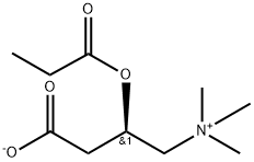
Propionyl-L-carnitine (120 mg/kg; p.o.; daily; starting immediately after aortic injury and lasting for 15 days) reduces the relative aortic intimal volume, increases apoptosis, up-regulates Bax without changing the Bcl-2 level, and reduces NF-κB, VCAM-1, MCP-1, and survivin in rats[3].
Propionyl-L-carnitine (30-120 mg/kg/day, p.o., drinking water) enhances wound healing and counteracts microvascular endothelial cell dysfunction in rats[4].
Propionyl-L-carnitine (3.6 mg/mL, infused into the renal artery at the rate of 0.4 mL/min) prevents renal function deterioration due to ischemia/reperfusion in male Sprague-Dawley rats[5].
Propionyl-L-carnitine (200 mg/kg/day, p.o., 4 weeks) corrects metabolic and cardiovascular alterations in diet-induced obese mice and improves liver respiratory chain activity[6].
Propionyl-L-carnitine (25? mg/kg, intrarectal, twice daily, for 1 week; 120 mg/kg, p.o., once daily, for 1 week) reduces intestinal damage and microvascular dysfunction in rat TNBS-induced acute and reactivated colitis[7].
Propionyl-L-carnitine (200 mg/kg, p.o., drinking water) increases NO synthesis by enhancing eNOS expression in spontaneously hypertensive rats[8].
Propionyl-L-carnitine (500 mg/kg, i.p., 10 successive days) prevents the progression of Cisplatin (HY-17394)-induced cardiomyopathy in a carnitine-depleted rat model[9].
Propionyl-L-carnitine (200 mg/kg per d, p.o., 20 weeks) reduces body weight and hyperinsulinaemia in obese Zucker rats[10].
| Animal Model: | Male Wistar rats (200-225 g, 13-14 weeks old), skin flap model and full-thickness skin wound model[4] |
| Dosage: | For the skin flap model, 100 mg/kg (dissolved in drinking water); for the full-thickness skin wound model, 30 mg/kg, 60 mg/kg, 120 mg/kg (dissolved in water). |
| Administration: | Oral gavage (p.o.), drinking water |
| Result: | Showed a faster blood flow recovery of the flap, an improved viability, reduced ROS production, increased NO level and iNOS expression (In the skin flap model).
Reduced the necrotic skin area after 8 days (in the skin flap model).
Had faster re-epithelialization, increased CD31+ capillaries, iNOS, VEGF and PlGF expression, and reduced Nox4+ microvessels (in the full-thickness skin wound model).
|