Polyethylene glycol immunogenicity: anti-polyethylene glycol antibody
Polyethylene glycol immunogenicity
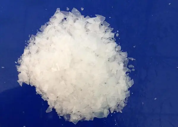
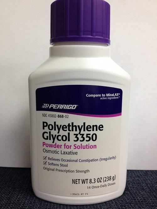
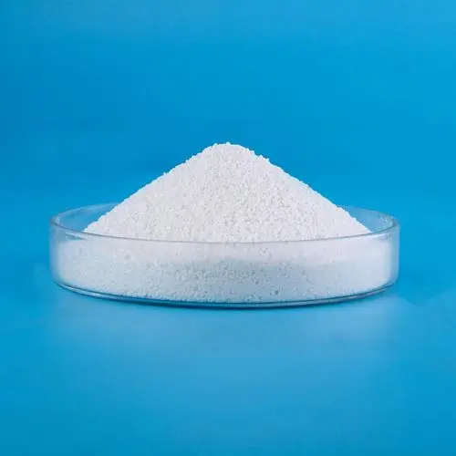
Polyethylene glycol (PEG) is a flexible, hydrophilic simple polymer that is physically attached to peptides, proteins, nucleic acids, liposomes, and nanoparticles to reduce renal clearance, block antibody and protein binding sites, and enhance the half-life and efficacy of therapeutic molecules. Some naive individuals have pre-existing antibodies that can bind to PEG, and some PEG-modified compounds induce additional antibodies against PEG, which can adversely impact drug efficacy and safety.
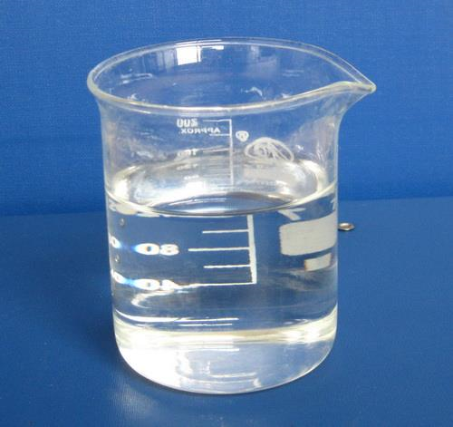
Attachment of polyethylene glycol (PEG) to small molecules, nucleotides, peptides, proteins, liposomes, and nanoparticles is widely used to improve their stability, solubility, and pharmacokinetic properties. Increased half-life in the circulation is particularly advantageous for injectable drugs because administration frequency can be reduced, leading to better patient compliance and quality of life. Due to the beneficial properties of PEG, a range of pegylated protein and non-protein medicines are clinically available, including RNA vaccines against SARS-CoV-2.
The size and number of PEG molecules attached to a compound can be varied depending on the desired purpose. A single linear or branched methoxy PEG (mPEG) molecule ranging in size from 12 to 60 kDa is attached to peptides, nucleotides, and small recombinant proteins to increase their hydrodynamic diameter, thereby reducing uptake by the kidney. On the other hand, multiple mPEG5000 molecules are attached to the surface of foreign enzymes to increase in vivo stability and block binding of anti-enzyme antibodies. Hundreds or even thousands of mPEG2000−lipid molecules are incorporated in liposomes and nanoparticles to reduce uptake by resident macrophages in the liver. Although PEG is typically depicted as a small linear molecule with dimensions on the order of a small protein, the actual contour lengths of commonly used PEG molecules range from 12.5 nm for PEG2000 to 253 nm for linear PEG40,000.
Pre-existing antibodies to PEG
Early studies using hemagglutination of PEG-modified red blood cells found between 0.2% and 25% of normal donors had antibodies specific to PEG in their plasma. In mice, a special population of B cells (B-1 cells) spontaneously secrete natural antibodies in the absence of exogenous immunization to provide pre-existing, immediate defense against microbial infections. The existence of analogous human B-1 cells is controversial, but other B cell populations may play a similar role in humans. A genomewide association study found that the presence of pre-existing anti-PEG IgM antibodies is associated with a specific variable segment of the immunoglobulin heavy-chain gene, suggesting that some individuals may have natural antibodies that bind PEG or are more sensitive to casual PEG exposure. Casual exposure to PEG compounds present in pharmaceutical, cosmetic, and health care products may induce anti-PEG antibodies,46 which may be promoted by inflammatory responses at sites of dermal abrasion and inflammation.
Anti-PEG antibody responses to thymus- dependent antigens
PEGylated proteins and peptides can elicit anti-PEG antibody responses by the classical T-cell-dependent pathway. Naive B cells express membrane-bound IgM and IgD immunoglobulins with the same antigen-binding specificity that act as B cell receptors (BCRs). B cells that express BCRs with specificity for PEG are activated when repeating epitopes present in the PEG backbone cross-link multiple BCRs. This can induce differentiation of naive B cells into plasmablasts that secrete IgM antibodies against PEG. However, a robust IgG antibody response requires that anti-PEG B cells receives additional signals or help from specialized CD4+ T cells called follicular helper T cells (TFH cells) in secondary lymphoid organs.
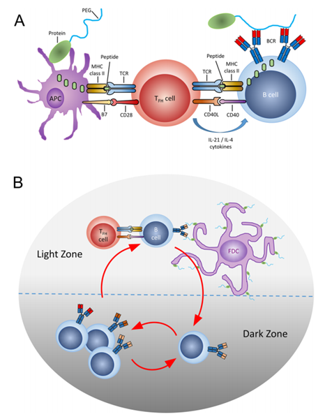
Thymus-dependent immune response against PEG. (A) Antigen-presenting cells take up pegylated proteins and present digested peptide fragments to activate specific TFH cells. Pegylated protein that is bound by anti-PEG immunoglobulins on PEG-specific B cells is digested and presented on MHC class II molecules to receive important signals from activated TFH cells (CD40L, IL-21, and other cytokines) to initiate somatic hypermutation of antibody variable region genes and class switch from IgM to IgG, IgA, or IgE in germinal centers. (B) Anti-PEG B cells undergo rapid proliferation and somatic hypermutation in the dark zone of a germinal center. B cells that display immunoglobulin with fortuitous mutations that increase PEG binding affinity can uptake greater amounts of pegylated protein from follicular dendritic cells (FDC) in the light zone. These B cells display sufficient peptide/MHC complexes to receive survival signals from TFH cells and recycle back to the dark zone for additional rounds of mutation and selection in a process called affinity maturation.
References:
[1] BING-MAE CHEN, Steve R R, Tian Lu Cheng. Polyethylene Glycol Immunogenicity: Theoretical, Clinical, and Practical Aspects of Anti-Polyethylene Glycol Antibodies[J]. ACS Nano, 2021, 15 9: 14022-14048. DOI:10.1021/acsnano.1c05922.
Related articles And Qustion
See also
Lastest Price from Polyethylene Glycol manufacturers
US $150.00/kg2025-11-21
- CAS:
- 25322-68-3
- Min. Order:
- 1kg
- Purity:
- 99%
- Supply Ability:
- 20tons

US $50.00-10.00/kg2025-09-02
- CAS:
- 25322-68-3
- Min. Order:
- 1kg
- Purity:
- 99%,Electronic grade(Single metal impurity≤ 100ppb) or pharmaceutical grade
- Supply Ability:
- 100kg

