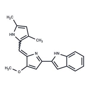
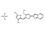
| In vitro | Obatoclax (GX15-070) inhibits several BCL-2 family proteins, with Ki values around 1-7 μM, demonstrating its potency in apoptosis regulation. In colorectal cancer cell lines (HCT116, HT-29, LoVo), it significantly reduces cell numbers in a dose- and time-specific manner, with the IC50 for cell proliferation at 72 hours being 25.85, 40.69, and 40.01 nM, respectively. At 400 nM for 24 hours, Obatoclax induces autophagy in OSCC cells and provokes an increase in G1-phase cell populations at doses of 50-200 nM for 24 hours. Similarly, it decreases cyclin D1 levels significantly at these concentrations. It leads to both phosphorylation-dependent and -independent cyclin D1 degradation in HCT116 and LoVo cells, with a notable decline in p-Cyclin D (T286) levels after treatment. Furthermore, Obatoclax inhibits GSK3β, activates p38 MAPK without significantly affecting ERK1/2 activity in HT-29 cells, and effectively inhibits the clonogenic potential of oral cancer cells across a range of concentrations (50-450 nM), underscoring its broader impact on cancer cell survival and proliferation mechanisms. |

 United States


