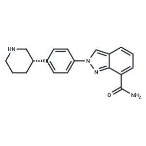
| Name | Niraparib |
| Description | Niraparib (MK-4827) is a PARP inhibitor that selectively targets PARP1 and PARP2 (IC50=3.8/2.1 nM), exhibiting antitumor activity, inhibiting DNA damage repair, and inducing apoptosis. |
| Cell Research | Proliferation assays were conducted in 96-well black viewplates, and 300 cells/well (250 cell/well for BRCA-1 wt) in culture medium, 190 μL/well (DMEM containing 10% FCS, 0.1 mg/mL penicillin-streptomycin, and 2 mM L-glutamine), were plated and incubated for 4 h at 37℃ under 5% CO2 atmosphere. Inhibitors were then added with serial dilutions, 10 μL/well to obtain the desired final compound concentration in 0.5% DMSO. The cells were then incubated for 7 days at 37℃ in 5% CO2 after which time viability was assessed. Briefly, with CellTiter-Blue solution prediluted 1:10 in medium, 100 μL/well was added and the cells left for 45 min at 37℃ under 5% CO2 and then a further 15 min at room temperature in the dark. The number of living cells was determined by reading the plate at fluorimeter, excitation at 550 nm and emission at 590 nm. Cell growth was expressed as the percentage growth with respect to vehicle treated cells. The concentration required to inhibit cell growth by 50% (CC50) was determined.(Only for Reference) |
| Kinase Assay | Enzyme assay is conducted in buffer containing 25 mM Tris, pH 8.0, 1 mM DTT, 1 mM spermine, 50 mM KCl, 0.01% Nonidet P-40, and 1 mM MgCl2. PARP reaction contains 0.1 μCi [3H]NAD+ (200?000 DPM), 1.5 μM NAD+, 150 nM biotinylated NAD+, 1 μg/mL activated calf thymus, and 1?5 nM PARP-1. Autoreactions utilizing SPA bead-based detection are carried out in 50 μL volumes in white 96-well plates. Compounds (e.g., MK-4827) are prepared in 11-point serial dilution in 96-well plate, 5 μL/well in 5% DMSO/Water (10× concentrated). Reactions are initiated by adding first 35 μL of PARP-1 enzyme in buffer and incubating for 5 min at room temperature and then 10 μL of NAD+ and DNA substrate mixture. After 3 h at room temperature, these reactions are terminated by the addition of 50 μL of streptavidin-SPA beads (2.5 mg/mL in 200 mM EDTA, pH 8). After 5 min, they are counted using a TopCount microplate scintillation counter. IC50 data is determined from inhibition curves at various substrate concentrations[1]. |
| In vitro | METHODS: The PDAC cell lines MIA-PaCa-2, PANC-1, Capan-1 and the OvCa cell lines OVCAR8, PEO1 were treated with Niraparib (0.1-200 µM) for 48 h, and the cells were assayed for viability using the CellTiter-Glo Luminescent Cell Viability Assay.
RESULTS: The IC50 of Niraparib on MIA-PaCa-2, PANC-1, Capan-1, OVCAR8, and PEO1 cells were 26 µm, 50 µm, 15 µM, 20 µM, and 28 µM, respectively.[1] [2].
METHODS: Ovarian cancer cells SKOV3 and UWB1.289 were treated with Niraparib (0.5-15 µM) for 48 h, and the expression levels of target proteins were detected by Western Blot.
RESULTS: Niraparib upregulated the expression of PD-L1 in SKOV3 and UWB1.289 cells. [2] |
| In vivo | METHODS: To detect anti-tumor activity in vivo, Niraparib (25 mg/kg, administered orally four times per week) and PD-L1 (10 mg/kg, administered intraperitoneally twice per week) were administered to C57BL/6 mice bearing ovarian cancer tumor ID8 for eight weeks.
RESULTS: Niraparib upregulated PD-L1 expression in ovarian tumors in vivo and synergized with PD-L1 blockade. [2]
METHODS: To detect anti-tumor activity in vivo, Niraparib (50 mg/kg, 0.5% methylcellulose) was administered by gavage to C57BL/6 mice bearing intracranial human-derived TNBC cell lines SUM149, MDA-MB-231Br, or MDA-MB-436 once daily for two weeks.
RESULTS: In the BRCA mutant MDA-MB-436 model, Niraparib increased median survival and decreased tumor load. However, it did not increase in the BRCA mutant SUM149 or BRCA wild-type MDA-MB-231Br models, despite high concentrations in intracranial tumors. [3] |
| Storage | Powder: -20°C for 3 years | In solvent: -80°C for 1 year | Shipping with blue ice/Shipping at ambient temperature. |
| Solubility Information | Ethanol : 60 mg/mL (187.27 mM), Sonication is recommended.
H2O : < 1 mg/mL (insoluble or slightly soluble)
10% DMSO+40% PEG300+5% Tween 80+45% Saline : 6 mg/mL (18.73 mM), Solution.
DMSO : 65.63 mg/mL (204.84 mM), Sonication is recommended.
|
| Keywords | V-PARP | TANK-1 | poly ADP ribose polymerase | PARP3 | PARP2 | PARP1 | PARP | ovarian | orally | Niraparib | MK4827 | MK 4827 | lung | Inhibitor | inhibit | DNA | damage | cancer | breast | bioavailable | Apoptosis | anti-tumor |
| Inhibitors Related | Stavudine | Aceglutamide | Urea | Cysteamine hydrochloride | Sodium 4-phenylbutyrate | Metronidazole | Citric Acid Triammonium | L-Methionine | Sodium butanoate | Dimethyl phthalate | Alginic acid | Dextran sulfate sodium salt (MW 5000) |
| Related Compound Libraries | Highly Selective Inhibitor Library | Failed Clinical Trials Compound Library | Bioactive Compound Library | Anti-Cancer Clinical Compound Library | Drug Repurposing Compound Library | Inhibitor Library | Anti-Cancer Approved Drug Library | FDA-Approved Drug Library | Anti-Aging Compound Library | Bioactive Compounds Library Max | Anti-Cancer Drug Library | Anti-Cancer Active Compound Library |

 United States


