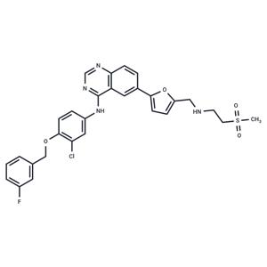
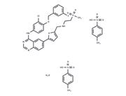
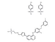
| Name | Lapatinib |
| Description | Lapatinib (GW572016) is an inhibitor of ErbB2 and EGFR (IC50=9.2/10.8 nM) with oral activity. Lapatinib has antitumor activity and can be used to treat advanced or metastatic breast cancer with HER2 overexpression. |
| Cell Research | Cells are plated in 96-well plates, in the media, at the following densities: HFF and HN5, 1000 cells/well and BT474, 5000 cells/well. After 24 h, the cells are exposed to vehicle (0.3% DMSO) or Lapatinib (1 nM, 10 nM, 100 nM, 1μM, 10μM, and 100μM). Lapatinib is removed from the cells after 72 h and is replaced by either DMEM containing 10% FBS and 50 μg/mL Gentamicin (HFF and HN5) or RPMI containing 10% FBS and 50 μg/mL Gentamicin (BT474). Methylene blue staining is performed at the time points over a total period of 16 days. Relative cell number is estimated using methylene blue staining. The absorbance at 620 nm is read in a Spectra microplate reader. Cell death and cell cycle analysis are assessed by propidium iodide staining and antibody detection of incorporated BrdUrd and staining with propidium iodide [1]. |
| Kinase Assay | The IC50 values for inhibition of enzyme activity are generated by measuring the inhibition of phosphorylation of a peptide substrate. The intracellular kinase domains of EGFR and ErbB2 are purified from a baculovirus expression system. EGFR and ErbB2 reactions are performed in 96-well polystyrene round-bottomed plates in a final volume of 45 μL. Reaction mixtures contain 50 mM 4-morpholinepropanesulfonic acid (pH 7.5), 2 mM MnCl2, 10 μM ATP, 1 μCi of [γ33P] ATP/reaction, 50 μM Peptide A [Biotin-(amino hexonoic acid)-EEEEYFELVAKKK-CONH2], 1 mM dithiothreitol, and 1 μL of DMSO containing serial dilutions of Lapatinib beginning at 10 μM. The reaction is initiated by adding the indicated purified type-1 receptor intracellular domain. The amount of enzyme added is 1 pmol/reaction (20 nM). Reactions are terminated after 10 minutes at 23°C by adding 45 μL of 0.5% phosphoric acid in water. The terminated reaction mix (75 μL) is transferred to phosphocellulose filter plates. The plates are filtered and washed three times with 200 μL of 0.5% phosphoric acid. Scintillation cocktail (50 μL) is added to each well, and the assay is quantified by counting in a Packard Topcount. IC50 values are generated from 10-point dose-response curves [1]. |
| Animal Research | CD-1 nude female mice are used for HN5 human tumor xenografts, which are initiated by injection of a cell suspension in PBS: Matrigel (1:1). C.B-17 SCID female mice are used for BT474 human tumor xenografts, which are initiated by implantation of tumor fragments (20-100 mg) from established tumors. Tumor cells and fragments are implanted by s.c. injection in the right flank. The s.c. tumors are measured with calipers, and mice are weighed twice weekly. Tumor weight is estimated from tumor volume using this formula: length×width2/2=tumor volume (mm3). Treatment begins when tumors are palpable, 3-5 mm in diameter. Lapatinib (30 and 100 mg/kg) is administered p.o. twice daily for 21 days in a vehicle of sulfo-butyl-ether-β-cyclodextrin 10% aqueous solution (CD10) [1]. |
| In vitro | METHODS: Fourteen human gastric and esophageal cancer cell lines were treated with Lapatinib (0.3125-10 µmol/L) for 6 days, and the cell counts were measured by particle counter.
RESULTS: N87, OE19, NUGC4, NUGC3, FU97, and SNU16 cells were sensitive to Lapatinib with IC50s of 0.01, 0.09, 0.35, 2.24, 4.86, and 8.58 μmol/L, respectively. [1]
METHODS: Human breast cancer cells MDA-MB-231 and SK-BR-3 were treated with Lapatinib (0.5-2 µM) for 48 h, and the expression levels of target proteins were detected by Western Blot.
RESULTS: The expression of PKM2 was significantly reduced in both MDA-MB-231 and SK-BR-3 cell lines treated with 1.0 µM Lapatinib compared to lower concentrations. [2] |
| In vivo | METHODS: To detect anti-tumor activity in vivo, Lapatinib (100 mg/kg) was administered intraperitoneally once daily for three weeks to CD-1 athymic nude mice bearing human gastric cancer tumor N87.
RESULTS: Lapatinib caused tumor regression in N87 xenografts. [1]
METHODS: To detect in vivo antitumor activity, Lapatinib (30-100 mg/kg administered by gavage twice daily for 21 days) and Topoteca (6-10 mg/kg administered intraperitoneally three times every four days) were administered by gavage to SCID mice harboring human mammary carcinoma tumor BT474.
RESULTS: The combination of Lapatinib and Topoteca showed enhanced efficacy in ER2+BT474 xenografts. [3] |
| Storage | Powder: -20°C for 3 years | In solvent: -80°C for 1 year | Shipping with blue ice/Shipping at ambient temperature. |
| Solubility Information | DMSO : 262.5 mg/mL (451.76 mM), Sonication is recommended.
H2O : < 1 mg/mL (insoluble or slightly soluble)
10% DMSO+40% PEG300+5% Tween 80+45% Saline : 9.3 mg/mL (16.01 mM), Solution.
Ethanol : < 1 mg/mL (insoluble or slightly soluble)
|
| Keywords | Lapatinib | Inhibitor | inhibit | HER2/ErbB2 | HER1 | GW-572016 | GW-2016 | GW2016 | GW 572016 | GW 2016 | GSK-572016 | GSK 572016 | Ferroptosis | ErbB-1 | Epidermal growth factor receptor | EGFR | Autophagy |
| Inhibitors Related | Stavudine | Cysteamine hydrochloride | Sodium 4-phenylbutyrate | Hydroxychloroquine | Guanidine hydrochloride | L-Glutamic acid | Paeonol | L-Cystine | L-Glutamine | Naringin | Alginic acid | Gefitinib |
| Related Compound Libraries | Bioactive Compound Library | Membrane Protein-targeted Compound Library | Tyrosine Kinase Inhibitor Library | Kinase Inhibitor Library | Anti-Cancer Clinical Compound Library | Drug Repurposing Compound Library | FDA-Approved Drug Library | FDA-Approved Kinase Inhibitor Library | Anti-Aging Compound Library | Heat-Clearing and Detoxifying Traditional Chinese Medicine Compound Library | Anti-Cancer Active Compound Library | Anti-Cancer Drug Library |

 United States


