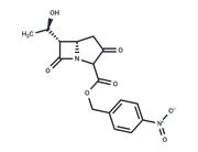
| Name | CFSE |
| Description | CFSE (CFDA-SE) is a fluorescent dye with cell membrane permeability. CFSE irreversibly binds to intracellular proteins in living cells and is used for the detection of cell proliferation. The labeled cells fluoresce in green color with excitation wavelength of 488 nm and emission wavelength of 518 nm. |
| Cell Research | Instructions:
I. Solution preparation
1. Preparation of mother solution: Take 1 mg CFDA-SE and dissolve it in 0.1794 mL DMSO to obtain 10 mM CFDA-SE mother solution.
Note: The mother solution is recommended to be stored at -20℃ or -80℃ away from light to avoid repeated freezing and thawing.
2. Preparation of working solution: Use pure DMEM to dilute the mother solution, usually with a concentration of 1-10μM.
Note: Please adjust the concentration of CFDA-SE working solution according to actual conditions.
II. Cell staining
1. Cell type:
1) Suspended cells: Centrifuge at 4℃, 1000 g for 3-5 minutes, discard the supernatant. Wash with PBS twice, 5 minutes each time.
2) Adherent cells: Discard the cell culture medium, add trypsin to dissociate the cells, and make a single cell suspension. Centrifuge at 4℃, 1000 g for 3-5 minutes, discard the supernatant. Wash with PBS twice, 5 minutes each time.
3. Add 1 mL CFDA-SE working solution and incubate at room temperature for 30 minutes.
4. Centrifuge at 400 g for 3-4 minutes at 4°C
5. Wash twice with PBS, 5 minutes each time.
6. Resuspend cells in serum-free cell culture medium or PBS and detect by fluorescence microscopy or flow cytometry. |
| In vitro | METHODS: 1 mL of cells and CFSE (5 μM in 110 μL PBS) were flipped and mixed to label the cells. CFSE-labeled CD8+OT-I T cells were cultured with dendritic cells pulsed with varying amounts of OVA for 3 days, and CFSE profiles were examined using Flow Cytometry.
RESULTS: CD8+ T cells divided 1-3 times according to the CFSE dilution peak, and more T cells divided at higher antigen concentrations. [1]
METHODS: Human erythroleukemia cell line K562, mouse lymphoma cell line YAC-1, human breast cancer cell line MCF-7, and human melanoma cell line A375 were treated with CFSE (1-10 μM) for 1-6 h. Cell death was detected by Flow Cytometry.
RESULTS: CFSE was non-toxic to the cells, as the cell death rate due to CFSE labeling was less than 5%. [2] |
| In vivo | METHODS: CFSE-labeled CD8+OT-I T cells were injected intravenously into the tail of C56BL/6J mice, followed by intravenous injection of OVA (20 μg), and CFSE profiles were measured three days later.
RESULTS: Most of the cells fell within 7 CFSE peaks, indicating that the cells had undergone up to 6 divisions. [1]
METHODS: To label thymocytes in vivo, CFSE (10 μM) was injected into the thymic lobes of anesthetized C56BL/6 mice.
RESULTS: CFSE labeled a representative sample of all thymocyte subpopulations and these cells migrated to peripheral lymphoid organs at a rate of approximately 2-3 x 10^6 cells/day. They enter the lymph nodes on day 1 post-injection and remain there for at least 21 days, while turnover is faster in the spleen. [3] |
| Storage | keep away from direct sunlight,store at low temperature | Powder: -20°C for 3 years | In solvent: -80°C for 1 year | Shipping with blue ice/Shipping at ambient temperature. |
| Solubility Information | Ethanol : < 1 mg/mL (insoluble or slightly soluble)
DMSO : 93 mg/mL (166.83 mM), Sonication is recommended.
H2O : < 1 mg/mL (insoluble or slightly soluble)
10% DMSO+40% PEG300+5% Tween-80+45% Saline : 3.3 mg/mL (5.92 mM), Sonication is recommeded.
|
| Keywords | Inhibitor | inhibit | CFSE |

 United States


