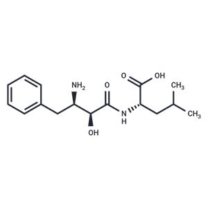
| Name | Bestatin |
| Description | Bestatin (Ubenimex) competitively inhibits many aminopeptidases, including B, N and leucine aminopeptidases. Ubenimex is a microbial metabolite and dipeptide with potential immunomodulatory and antitumor activities. Aminopeptidases has been implicated in the process of cell adhesion and invasion of tumor cells. Therefore, inhibiting aminopeptidases may partially attribute to the antitumor effect of ubenimex. This agent also activates T lymphocyte, macrophage, and bone marrow stem cell as well as stimulates the release of interleukin-1 and -2, thus further enhances its antitumor activity. |
| Cell Research | Growing cells (1×10⁶ to 2×10⁶ cells/mL) are diluted to 1.0×10³ cells/mL and transferred (3 mL) into a well in a 12-well multiwell plate (2.5-cm diameter/well). Cells are treated with 0, 10, 50, 100, 300, or 600 μM Bestatin and allowed to grow at 21°C shaking at 180 rpm for 48 h. A hemocytometer is used to measure cell density after 0, 24, and 48 h. |
| Kinase Assay | Cells are harvested, washed, and lysed in NP-40 lysis buffer (50 mM Tris-HCl [pH 7.5], 150 mM NaCl, 0.5% NP-40). Total cell protein is quantified using the Bradford assay and 1-mg/mL protein aliquots are made. Ten microliters of total cell protein is mixed with 290 μL of substrate solution (0.1 mg/mL dithiothreitol [DTT], 0.1 mg/mL albumin, and 1 mM alanine-β-naphthylamide). Fluorometric measurements (340 nm excitation, 400 nm emission) are made after 15 and 30 min. The slope of the line between the 15- and 30-min measurements is used to represent aminopeptidase activity. Total cell protein is preincubated with bestatin, amastatin, puromycin, EDTA, and/or ZnCl2 for 20 min before the fluorometric aminopeptidase assay. |
| In vitro | In mice bearing B16-BL6 melanoma tumors on their dorsolateral sides, Bestatin reduced the number of blood vessels in established primary tumor masses. Additionally, Bestatin significantly inhibited angiogenesis induced by melanoma cells in the dorsal air sac assay in mice. In EGDA rats, Bestatin decreased the incidence of EAC from 57.7% to 26.1%. Moreover, Bestatin demonstrated statistically significant inhibition of leukotriene B4 biosynthesis in the esophageal tissues of EGDA rats. |
| In vivo | Bestatin exhibits a concentration-dependent inhibition of SN12M cell invasion into reconstituted basement membrane (matrigel). It also concentration-dependently inhibits the degradation of type IV collagen by tumor cells in a non-neoplastic condition medium. In U937 cells, bestatin enhances the activity of caspase-3, inducing DNA laddering and fragmentation. Additionally, in SN12M, bestatin inhibits the hydrolytic activity of aminopeptidase substrates. Furthermore, bestatin inhibits tube formation in human umbilical vein endothelial cells. Through binding to leucine aminopeptidase fixed on the cell surface, bestatin directly stimulates lymphocytes (and monocytes), whereas it indirectly does so by impeding the degradation metabolism of the phagocytosis stimulating hormones through the inhibition of aminopeptidase B. |
| Storage | Powder: -20°C for 3 years | In solvent: -80°C for 1 year | Shipping with blue ice. |
| Solubility Information | 1eq. NaOH : 15.4 mg/mL (50 mM)
|
| Keywords | Aminopeptidase | Bacterial | Inhibitor | Antibiotic | Bestatin | inhibit |
| Related Compound Libraries | Anti-Tumor Natural Product Library | Bioactive Compound Library | Anti-Cancer Clinical Compound Library | Natural Product Library | Anti-Viral Compound Library | Drug Repurposing Compound Library | Microbial Natural Product Library | Natural Product Library for HTS | Anti-Cancer Active Compound Library | Anti-Cancer Drug Library |

 United States


