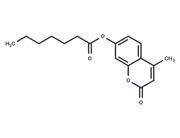
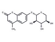
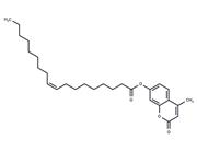
| Name | 4-Methylumbelliferyl phosphate |
| Description | 4-Methylumbelliferyl phosphate (4-MUP) (4-MUP) is used as a fluorogenic substrate of alkaline phosphatases. |
| Cell Research | I. Alkaline phosphatase activity detection
1. Preparation of 4-MUP solution: Dissolve 4-MUP in an appropriate buffer, such as Tris-HCl (pH 8.0), with a concentration generally between 0.1-1 mM.
2. Add sample: Add a sample containing alkaline phosphatase (such as serum or cell lysate) to the 4-MUP solution. Adjust the final reaction volume according to the desired sensitivity.
3. Incubate the reaction system at 37°C, usually for 30 minutes to 1 hour, to allow the enzyme to dephosphorylate 4-MUP.
4. Fluorescence measurement: After incubation, measure the fluorescence of 4-methylumbelliferone by a fluorescence spectrophotometer or microplate reader, with an excitation wavelength of 360 nm and an emission wavelength of 450 nm.
5. Data analysis: The fluorescence intensity is proportional to the activity of alkaline phosphatase. By comparing with the standard curve, the enzyme activity in the sample can be quantified.
II. Application in Enzyme-Linked Immunosorbent Assay (ELISA)
1. Plate coating: Coat the antigen or antibody of interest on the microplate.
2. Add alkaline phosphatase-labeled antibody: Add the antibody or enzyme-labeled probe linked to alkaline phosphatase.
3. Add substrate: Add 4-MUP substrate after washing.
4. Fluorescence detection: After incubation, measure the fluorescence signal using a microplate reader or fluorescence plate reader with an excitation wavelength of 360 nm and an emission wavelength of 450 nm.
5. Result interpretation: Quantify the amount of antigen or antibody based on the fluorescence intensity.
III. Alkaline phosphatase detection in cell culture
1. Cell treatment: Culture cells in appropriate culture medium and treat as needed.
2. Add 4-MUP: Add 4-MUP solution to the cell culture medium.
3. Fluorescence monitoring: After incubation, use a fluorescence microscope or microplate reader for fluorescence measurement.
4. Quantitative analysis: Quantify enzyme activity by fluorescence intensity to evaluate changes in alkaline phosphatase during treatment or differentiation.
IV. Alkaline phosphatase detection in tissue sections
1. Tissue section preparation: Prepare tissue sections using conventional histological methods.
2. Add 4-MUP: Soak the sections in 4-MUP solution, usually incubated at 37°C.
3. Fluorescence microscopy observation: After incubation, use a fluorescence microscope to observe the fluorescence signal.
5. Analysis: Evaluate the activity of alkaline phosphatase by observing the fluorescence distribution and intensity in the tissue. |
| In vitro | 4-Methylumbelliferyl phosphate has a significant advantage over currently available substrates in that its useful concentration range extends to about 0.1 μM. If the concentration of 4-Methylumbelliferyl phosphate is held constant and the pH is varied, the activity of the enzyme passes through a maximum. |
| Storage | keep away from direct sunlight,store at low temperature | Powder: -20°C for 3 years | In solvent: -80°C for 1 year | Shipping with blue ice/Shipping at ambient temperature. |
| Solubility Information | H2O : 20 mg/mL (78.07 mM), Sonication is recommended.
|
| Keywords | substrate | phosphatases | Phosphatase | MUP | kinetic | Inhibitor | inhibit | fluorogenic | Alkaline | 4-Methylumbelliferyl Phosphate | 4Methylumbelliferyl phosphate | 4-Methylumbelliferyl | 4 Methylumbelliferyl phosphate |
| Inhibitors Related | PTP1B-IN-22 | Ellagic acid | β-Glycerophosphate disodium salt pentahydrate | L-Ascorbic acid 2-phosphate trisodium | Idoxuridine | Tartaric acid disodium dihydrate | Cyclosporine | Cis-5-Norbornene-exo-2,3-dicarboxylic Anhydride | Stearic acid | CaMKP Inhibitor | Cyclosporin A | β-Glycerophosphate disodium salt hydrate |
| Related Compound Libraries | Human Metabolite Library |

 United States


