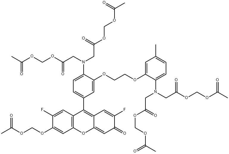
Fluo-4 AM is a cell-permeable Ca2+-indicator that is metabolized by intracellular esterase, leading to a bright green fluorescent signal upon Ca2+-binding (excitation/emission λ at 494/506 nm). Fluo-4 AM is used for visualization and measurement of intracellular Ca2+. It is well suited for fluorometric and imaging applications such as microscopy, flow cytometry, spectrofluorometry, and fluorometric high-throughput microplate screening assays [1].
Fluo-4 AM is similar in structure and spectral properties to the widely used Ca2+-indicator, Fluo-3, but it has certain advantages over Fluo-3, such as brighter fluorescence emission, high rate of cell permeation, and a Kd for Ca2+ in buffer of 345 nM. Because of its higher fluorescence emission intensity, Fluo-4 AM can be used at lower intracellular concentrations, making its use less toxic for live cells.
As Fluo-4 AM does not covalently bind to cellular components, it may be actively effluxed from the cell by organic anion transporters. In vivo cell imaging with Fluo-4 AM is usually performed within one or two hours after loading, but the dye can be re-loaded to cells if it is needed. Fluo-4 AM can also be fixed in situ by EDC/EDAC for downstream immunofluorescence studies.
Fluo-4 AM has low solubility in the water. It is recommended to prepare 1 mM stock solution in labeling grade DMSO prior to cell loading. Use the final concentration of 1-5 μM and incubation at 37 °C for 15-60 min as a start point of your protocol.