ANTI-PD-L1抗体
- CAS号:
- 英文名:Anti-PD-L1 Antibody
- 中文名:ANTI-PD-L1抗体
- CBNumber:CB15717168
- 分子式:
- 分子量:0
- MOL File:Mol file
- 生物来源 :rabbit
ANTI-PD-L1抗体性质、用途与生产工艺
- 描述 兔单克隆抗体[28-8] to PD-L1
- 宿主 Rabbit
- 来源 Monoclonal Rabbit IgG Clone #015
- 偶联物 Unconjugated
- 制备 This antibody was obtained from a rabbit immunized with purified, recombinant Human PD-L1 / B7-H1 / CD274 (rh PD-L1 / B7-H1 / CD274; Catalog#10084-H08H; NP_054862.1; Met 1-Thr 239).
- 经测试应用 适用于: IHC-P, WB, Flow Cyt, ICC/IF
-
品牌示例
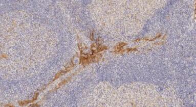
Immunohistochemistry (Formalin/PFA-fixed paraffin-embedded sections) - Anti-PD-L1 antibody [28-8] (ab205921)

IHC image of ab205921 staining PD-L1 in human tonsil formalin fixed paraffin embedded tissue sections*, performed on a Leica BOND RX (Polymer Refine kit). The section was pre-treated using heat mediated antigen retrieval with EDTA buffer (pH9, epitope retrieval solution 2) for 30 mins at 98°C. The section was then incubated with ab205921, 5μg/ml working concentration, for 60 mins at room temperature and detected using an HRP conjugated compact polymer system for 8 minutes at room temperature. DAB was used as the chromogen for 10 minutes at room temperature. The section was then counterstained with hematoxylin, blued, dehydrated, cleared and mounted with DPX.
For other IHC staining systems (automated and non-automated) customers should optimize variable parameters such as antigen retrieval conditions, primary antibody concentration and antibody incubation times.
*Tissue obtained from the Human Research Tissue Bank, supported by the NIHR Cambridge Biomedical Research Centre - 背景 Programmed death-1 ligand-1 (PD-L1, CD274, B7-H1) has been identified as the ligand for the immunoinhibitory receptor programmed death-1(PD1/PDCD1) and has been demonstrated to play a role in the regulation of immune responses and peripheral tolerance. PD-L1/B7-H1 is a member of the growing B7 family of immune molecules and this protein contains one V-like and one C-like Ig domain within the extracellular domain, and together with PD-L2, are two ligands for PD1 which belongs to the CD28/CTLA4 family expressed on activated lymphoid cells. By binding to PD1 on activated T-cells and B-cells, PD-L1 may inhibit ongoing T-cell responses by inducing apoptosis and arresting cell-cycle progression. Accordingly, it leads to growth of immunogenic tumor growth by increasing apoptosis of antigen specific T cells and may contribute to immune evasion by cancers. PD-L1 thus is regarded as promising therapeutic target for human autoimmune disease and malignant cancers.
-
检测原理
双抗体夹心法测定标本中PD-L1水平。用纯化的PD-L1抗体包被微孔板,制成固相抗体,往包被单抗的微孔中依次加入PD-L1,再与HRP标记的PD-L1抗体结合,形成抗体-抗原-酶标抗体复合物,经过彻底洗涤后加底物TMB显色。TMB在HRP酶的催化下转化成蓝色,并在酸的作用下转化成最终的黄色。颜色的深浅和样品中的PD-L1呈正相关。用酶标仪在450nm波长下测定吸光度(OD值),通过标准曲线计算样品中PD-L1浓度。
ANTI-PD-L1抗体是一类可以特异性结合PD-L1的多克隆抗体,主要用于检测PD-L1的Western Blot、IHC-P、IF、ELISA、Co-IP等多种免疫学实验。 -
PD-L1
PD-1的全称是程序性死亡受体-1,属于CD28超家族成员,是由268个氨基酸组成的I型跨膜蛋白。它是在1992年由日本京都大学的Tasuku Honjo教授发现的,可在T细胞、B细胞等免疫细胞表面表达。不过在T细胞未活化时,几乎不表达PD-1,仅在T细胞活化之后,PD-1才在T细胞表面表达。
PD-L1(programmed death ligand 1)全称程序性死亡受体配体1,是PD-1(programmed cell death 1,程序性死亡受体1)的配体。PD-L1属于细胞膜上的一个跨膜蛋白,表达在T细胞、B细胞等免疫细胞以及肿瘤细胞上,研究表明当肿瘤细胞膜上的PD-L1与T细胞等免疫细胞上的PD-1结合后,肿瘤细胞发出抑制性信号,进而T细胞不能识别肿瘤细胞和对肿瘤细胞产生杀伤作用,机体的免疫功能受到抑制。
- 公司名称:武汉佰乐博生物技术有限公司
- 联系电话:027-65279366 18108604356
- 电子邮件:products@biolabreagent.com
- 国家:中国
- 产品数:9868
- 优势度:58
- 公司名称:北京百普赛斯生物科技股份有限公司
- 联系电话: 18514007688
- 电子邮件:jiaxin.zhao@acrobiosystems.com
- 国家:中国
- 产品数:1264
- 优势度:58
- 公司名称:Enzo Biochem Inc
- 联系电话:80-Enzo Biochem Inc. 18009420430
- 电子邮件:info-usa@enzolifesciences.com
- 国家:中国
- 产品数:124812
- 优势度:58
- 公司名称:西格玛奥德里奇(上海)贸易有限公司
- 联系电话:(+86) 21 2033 8288
- 电子邮件:sigmaname@qq.com
- 国家:中国
- 产品数:292394
- 优势度:58
