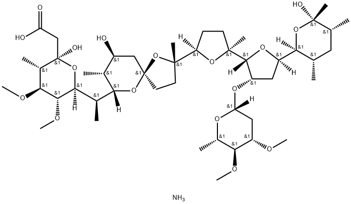
Maduramycin ammonium salt is a white crystalline powder with a melting point of 165-167°C. It is insoluble in water and soluble in organic solvents. Because of its broad anticoccidial spectrum and not easy to develop drug resistance, it is widely used. The dosage is very small. At present, our country stipulates that the amount of maduramycin ammonium salt added to the feed for preventing chicken coccidiosis is 5mg/kg.
Polyether antibiotic chemically related to the lonomycins. Coccidiostat
Maduramicin ammonium is prepared from maduramicin by taking advantage of the acidic carboxylic acid which ionises and readily forms the salt in ammonium hydroxide solutions. The ammonium salt is the preferred formulation in animals to prevent coccidiosis and to promote growth. Maduramicin is also active against Treponema and Cryptosporidium. Maduramicin is an ionophore, forming complexes with monovalent cations, with a higher affinity for K+ than Na+.
Maduramcin ammonium is prepared from maduramicin by taking advantage of the acidic carboxylic acid which ionises and readily forms the salt in ammonium hydroxide solutions. The ammonium salt is the preferred formulation in animals to prevent coccidiosis and to promote growth. Maduramicin is also active against Treponema and Cryptosporidium. Maduramicin is an ionophore, forming complexes with monovalent cations, with a higher affinity for K+ than Na+.
ChEBI: Maduramicin is a glycoside.
| Animal Model: | ICR mouse (Maduramicin-induced rhabdomyolysis model)[3] |
| Dosage: | 3.5, 7 mg/kg |
| Administration: | Oral gavage (p.o.), once daily for 7 days |
| Result: | Significantly reduced body weight, increased AST, ALT, LDH, BUN, and CK levels;
Caused that liver exhibited vacuolar degeneration, kidney showed renal tubular epithelial cell shedding, and myocardial and skeletal muscle fibers were disorganized and partially lysed. |
| Animal Model: | Wistar rats (Maduramicin-induced skeletal muscle injury model) |
| Dosage: | 1.75, 3.5, 7 mg/kg |
| Administration: | Oral gavage (p.o.), once daily for 7 days |
| Result: | Dose-dependently increased LC3-II and p62 protein levels in skeletal muscle, indicating impaired autophagic flux;
Upregulated eIF2α phosphorylation and ATF4 expression, activating endoplasmic reticulum stress;
Increased AMPK phosphorylation, suggesting activation of the AMPK signaling pathway. |