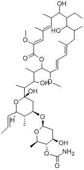
Description
Concanamycin A (80890-47-7) is a potent and specific inhibitor of the vacuolar (V-type) H+-ATPase which can induce apoptotic cell death in various cell lines.1,2Inhibits cell surface expression of virus envelope glycoproteins.3Dramatically increases the rate of extracellular vesicle release from a variety of cell types.4Inhibits autophagy by blocking lysosomal acidification.5
Uses
Concanamycin A has been used:
- as a lysosomal inhibitor in young and old fibroblasts
- as a vacuolar-type H+-ATPase inhibitor in presynaptic vesicles
- as a lysosomal acidification blocker in HepG2 hepatocytes cells
Uses
Concanamycin A is the major analogue of the concanamycin complex produced by Streptomyces sp.. It has been shown to act as a potent and specific vacuolar-ATPase inhibitor. Concanamycin A inhibits the acidification of organelles and blocks cell surface expression of viral envelope glycoproteins without affecting their synthesis. It also interferes with intracellular protein trafficking and inhibits perforin- and Fas-based lytic pathways in cell-mediated cytotoxicity. Concanamycins are structurally related to the bafilomycins.
Definition
ChEBI: A concanamycin in which the lactone ring contains 4 double bonds and is substituted by 4 methyl groups, 2 hydroxy groups, 2 methoxy groups and an ethyl group.
General Description
Chemical structure: macrolide
Biological Activity
Specific inhibitor of V-type (vacuolar) H + -ATPase that displays > 2000-fold selectivity over other H + -ATPases (IC 50 values are 9.2, > 20000, > 20000 and > 20000 nM for yeast V-type, F-type, P-type H + -ATPases and porcine P-type Na + ,K + -ATPase respectively). Blocks cell surface expression of virus envelope glycoproteins without affecting synthesis and exhibits cytotoxicity in several cell lines.
Biochem/physiol Actions
Concanamycin A (ConA) inhibits acidification of organelles and perforin-mediated cytotoxicity. It is a vacuolar-type v-ATPase inhibitor. ConA possesses antiprotozoal and antineoplastic properties. It mediates inhibition of the negative factor (Nef) protein of the human immunodeficiency virus.
Background
Concanamycin A is a macrolide antibiotic derived from S. diastatochromogenes that is effective against several fungi and yeasts. Concanamycin A is an exceptionally potent and specific inhibitor of the ATP-driven proton pumps known as vacuolar type H+-ATPases. V-ATPases acidify intracellular compartments and translocate protons across the plasma membrane. Intracellular V-ATPases play an important role in endocytosis and intracellular membrane trafficking, while plasma membrane V-ATPases are important in processes such as urinary acidification and bone resorption. Treatment of murine cells with Concanamycin A results in apoptosis, evidenced by an increase in fragmented DNA and the number of apoptotic cells with hypodiploid DNA. Concanamycin A induced production of nitric oxide and decreased cell growth and survival in mouse leukemic monocyte cells. Concanamycin A reversed the downregulation of cell surface MHC-I by the HIV-encoded accessory protein Nef, suggesting a possible therapeutic role of Concanamycin A in enhancing the immune-mediated clearance of HIV-infected cells.
References
[1] NISHIHARA T. Specific Inhibitors of Vacuolar Type H+-ATPases Induce Apoptotic Cell Death[J]. Biochemical and biophysical research communications, 1995, 212 1: Pages 255-262. DOI:
10.1006/bbrc.1995.1964[2] JANGJA HONG. Nitric oxide production by the vacuolar-type (H+)-ATPase inhibitors bafilomycin A1 and concanamycin A and its possible role in apoptosis in RAW 264.7 cells.[J]. Journal of Pharmacology and Experimental Therapeutics, 2006, 319 2: 672-681. DOI:
10.1124/jpet.106.109280[3] M MUROI. Folimycin (concanamycin A), a specific inhibitor of V-ATPase, blocks intracellular translocation of the glycoprotein of vesicular stomatitis virus before arrival to the Golgi apparatus.[J]. Cell structure and function, 1993, 18 3: 139-149. DOI:
10.1247/csf.18.139[4] ANIL G CASHIKAR P I H. A cell-based assay for CD63-containing extracellular vesicles.[J]. ACS Applied Bio Materials, 2019: e0220007. DOI:
10.1371/journal.pone.0220007[5] SYLWIA GRADZKA. Inhibitor of apoptosis proteins are required for effective fusion of autophagosomes with lysosomes.[J]. Cell Death & Disease, 2018: 529. DOI:
10.1038/s41419-018-0508-y